| Product name | Soft tissue liposarcoma round cell |
| Cat. No. | 1X88503B |
| No. of samples | 1 |
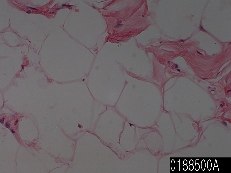
| Description | soft tissue, liposarcoma, round cell 51 years, female Age/Sex : 51/F |
| Price | 197 EUR |
| 260 USD | |
| 170 GBP |
 Product Related Literature
Round cell liposarcoma, sarcoma bond is a form of discrimination mucus liposarcoma is an insufficient low-grade. It is unclear, of cellular components round myxosarcoma result in a typical this is not the case in tumors behave as sarcoma how high. Twenty nine cases limb mucus liposarcoma, it is necessary to affect the adverse prognosis examined the size of the round cell components with or without components semiquantitate fat sarcoma round cells. Estimate of the proportion of the transition zone necrosis, round cell sarcoma, and myxosarcoma is obtained for each slide in all cases. It was defined as one that showed the cells increased compared with myxoid liposarcoma typical, however, transition zone remains a distinction plexiform vascular pattern spindled cells remain easy, and there was no boundary overlap of nuclear are out. It is subtracted from the total available material for evaluating the amount of necrosis, a composite measure of the proportion of the transition zone round cells and mucus is obtained.
(Median 14 cm) was located in the upper portion 27 of the lower limbs, the size of the tumor is different FRM 3 ~ 30 cm tumor of two. 29 and mucus components and 100% range 12, all tumors were (mean 73%). Range of transition element to the tumor of 29 of all is (11% average) 88 percent from 0. (To 88 percent range from 4) twenty one tumor, the transition region. (Average, 0%) range of circular cage component for all 29 tumors was 58 percent from 0. (58% range from 1) twelve tumor, round cell area. I died because of unrelated 202 months (median 96 months) from 24, or a disease-free patients Seventeen is alive. 12 cases died of disease (median, 53 months) in 180 months or 10, or was alive one of the transition. Patients to the original tumor having> round cell component of 5%, with a higher incidence of death and significant transition as a result of the disease compared with (P = 0.05) round cell liposarcoma or 5% = < had. In addition, only patients with mucus liposarcoma with a transition zone to perform worse compared with mucus liposarcoma. In conclusion, we> Passive Cell 5% were found to predict high risk of death or metastasis from disease. In addition, the transition zone does not appear that you want to change the forecast of only mucus liposarcoma. Therefore, those areas, must be specified as a class only liposarcoma of round cells clearly.
Even before undifferentiated sarcoma polymorphism (abbreviated PUS), of (abbreviation MFH), undifferentiated sarcoma polymorphism, malignant fibrous histiocytoma is a type of soft tissue sarcoma. It is considered a diagnostic exclusion of sarcoma, which can be classified more accurately. Depending on the histological subtype, variable behavior is (see LS, this term) type of position on a limb liposarcoma mainly round / mucus cell liposarcoma (MRCLS). For round cells and mucus is a histological subtype of different LS. MRCLS presents at a young age than the LS of other subspecies and to 35-55 year-old age typical of diagnosis. It occurs in the extremities (usually thigh) Mainly, this does not occur in subcutaneous tissue or rare retroperitoneal, many. One-third of the cases MRCLS is the metastatic spread of soft tissue area with metachronous multifocal distribution or synchronization of fat surface area of axillary retroperitoneal, trunk, and pericardium and abnormal bone.
MRCLS lesions of 90% has lead to fusion of DDIT3 16p11 gene on chromosome 12q13 and the region with characteristic chromosomal translocation FUS. As a result, dysregulating, hence the creation of FUS-DDIT3 fusion protein that promotes malignant transformation of dysregulating fat cell differentiation, cell cycle control of RNA transcription. Table is established, is carried out magnetic resonance imaging (CT) or a (MRI) computed tomography. Lesions of the chest and abdomen do not need a biopsy before as long as possible resection is very painful incomplete or not high. Normally, in order to identify the stage of disease and (mucus round the cellular component) main histological subtype, limb lesions take a sample from multiple biopsies. Rounding single, elliptical, MRCLS tumors are made up of prominent mucus between quality and small signet ring lipoblasts plexiform container and nonlipogenic primitive mesenchymal cells. The diagnosis can be confirmed by evidence DDIT3, FUS movement of fluorescence in RT-PCR in situ hybridization, or (FISH). Predict a poor prognosis and to determine tumor of high-level parts of the circular cell to determine whether it is a mucus <(5% round cell component) round cell (or 5% of the cell round component)>.
The MRCLS, you may be confused Ewing sarcoma, (see these terms) undifferentiated sarcoma polymorphism and lymphoma. Mucus other tumors should also be excluded. Treatment includes the surrounding tissue for mucus of low-grade liposarcoma and surgical resection of the tumor. In rare cases, amputation is required. Tumors can be treated with radiation therapy of preoperative or postoperative chemotherapy and / or preoperative. Compared to other subtypes, high-grade LS circuit cell MRCLS is resectable or slightly large (> 5 CM) respond well to treatment with radiation therapy or chemotherapy. Ecteinascidin while being used as second-line treatment, usually, and ifosfamide chemotherapy drug doxorubicin is a treatment option in the first row. Monitoring lifelong It is recommended that you monitor the recurrence and distant metastasis primary site. The prognosis of good MRCLS for patients with (measured as less than 5% of the cell round component and pure mucus) mucus of low-grade liposarcoma, 5-year survival rate is 92%. (5% or more) in the 5-year survival rate of 74%, round cell component is associated with poor prognosis very important.