| Product name | Kidney chronic pyelonephritis |
| Cat. No. | 7143000A |
| No. of samples | 1 |
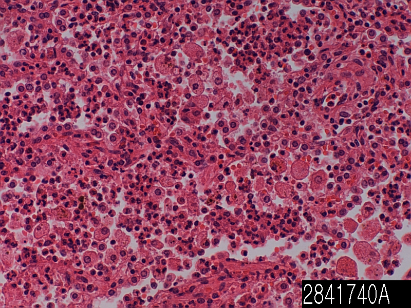
| Description | kidney chronic pyelonephritis Age/Sex : 49/M |
| Price | 197 EUR |
| 260 USD | |
| 170 GBP |

Product Related Literature
Kidney chronic pyelonephritis ascending urinary tract infections pyelonephritis, has reached the pelvis of the kidney or pyelum. It is a form of nephritis with pyelitis. Severe cases of pyelonephritis, there is a possibility that (collection of pus around the kidney) pyonephrosis (systemic inflammatory response to infection) urinary tract sepsis, renal failure, cause even death. Back to the costovertebral angle on the side affected, to tenderness and nausea, urination with radiant heat, rapid heart beat, pain, pyelonephritis, exhibit abdominal pain. Pyelonephritis can be accompanied by rapid breathing, low blood pressure, severe shaking, such as delirium, sometimes signs of septic shock by proceeding urinary tract sepsis. to antibiotic therapy, surgery and ureteroscopy, as percutaneous renal nefrostomiya percutaneously or sometimes, to prevent its recurrence, pyelonephritis and require treatment of the root cause of any. Pyelonephritis Xanthogranulomatous, is a rare form of chronic pyelonephritis, (removal of kidney) renal is necessary for final processing in general here.
Most cases of “community-acquired” pyelonephritis are due to bowel organisms into the urinary tract. Biological general is enterococci and Escherichia coli (70-80%). Enterococci and Escherichia coli, as well as caused by bacteria, nosocomial infection (for example, various types of Klebsiella and Pseudomonas aeruginosa) that allows other organisms, rare in society. Most cases of pyelonephritis, start lower urinary tract, prostatitis and cystitis in particular. It is possible to be able to mature E. coli biofilm to form intracellular bacterial communities to (the IBC), and to penetrate the surface umbrella cells of the bladder. If there is resistance to the response of the immune system and antibiotics, biofilms of these producing E. coli, provides the instructions, such as pyelonephritis, the potential for recurrent urinary tract infection. Risk is increased in the following situations:.
If the kidney stone is suspected, kidney, ureter, bladder, and the impermeable radiation (eg, based on the presence or absence of an imbalance of blood or urine pain characteristics colic) X-ray (KUB film) it is possible to help to identify the stone. With the exception of the stone very unusual consisting of drug residues of certain urine, diagnostic modality of choice in the X-ray evaluation of nephrolithiasis.All stones 5 mm section noncontrast spiral CT suspected and, if possible, detected by CT scan is possible. In patients with recurrent rise urinary tract infections, it may be necessary to exclude polycystic kidney disease or reflux vezikoureteralen of anatomical abnormality. The test used in this setting, ultrasound examination of voiding cystourethrography and kidney are included. The abdominal ultrasonography and CT, are useful in the diagnosis of xanthogranulomatous pyelonephritis, to obtain the serial image to distinguish the status of kidney cancer may be useful.
Are line antibiotic susceptibility tests and suspected urine culture and nephritis in a patient, and is adjusted based on the pathogenic bacterium primary care. In most cases of nephritis due to bacterial infection, which is the mainstay of antibiotic therapy. Depends on the antibiotic susceptibility profiles and type of soiling of the body and trimethoprim / sulfamethoxazole fluoroquinolones, cephalosporins, and aminoglycosides, alone or selection of antibiotics, can include combinations.
Is a low prevalence of resistant bacteria in patients who do not require hospitalization, oral fluoroquinolones such as levofloxacin or ciprofloxacin, is an appropriate choice for the initial treatment. In areas where there is a high fluoroquinolone resistance rate, that continuation of treatment with oral fluoroquinolones, starting treatment, such as an aminoglycoside or a single intravenous dose, long-acting antibiotic ceftriaxone then is useful it, and. If it uropathogen susceptible are known, Oral trimethoprim / sulfamethoxazole is a suitable choice for treatment. When using the trimethoprim / sulfamethoxazole when the sensitivity is not known, it is possible to start processing the aminoglycoside, or a single intravenous dose of long-acting antibiotic ceftriaxone is useful . The oral β-lactam antibiotics, efficiency is lower than other means that can be used for the treatment of pyelonephritis.
Usually, has been admitted to the hospital for intravenous antibiotics and intravenous hydration people with acute pyelonephritis accompanied by fever and leukocytosis. Treatment begins carbapenem and cephalosporin intravenous fluoroquinolones, aminoglycosides, or spectrum penicillin, broad in general. Often, a combination therapy of antibiotics, have been used in such a situation. The treatment regimen was selected based on the sensitivity of the profile (s) a particular pathogen and local resistance data.
In the course of antibiotic treatment, I have closely monitored the serial number of the temperature and white blood cells. Patient, until 24 to 48 hours afebrile least, continue the intravenous antibiotics typically then antibiotics equivalent, may be all the period administration of the treatment two weeks. Ingestion vacuum infusion, it is possible dead loss (by raising the temperature), and is administered to compensate for the diuretic and optimization vasodilation. Percutaneous ureteral stent placement or Nefrostomiya may be given to relieve disorders caused by the stone. Children with acute pyelonephritis can be treated effectively short course of intravenous therapy (cefixime, amoxicillin / clavulanate and Ceftibuten) or oral antibiotics (2 to 4) followed by oral therapy. If intravenous treatment is selected, a single daily dose of aminoglycosides is safe and effective. Treatment of xanthogranulomatous pyelonephritis, including surgery and antibiotics. Is effective for some people with the disease (partial nephrectomy) has been established polarity resection but nephrectomy is a surgical treatment of the best in most cases. Observation with serial imaging, may be appropriate in unusual circumstances.