| Product name | Placenta |
| Cat. No. | 0088100B |
| No. of samples | 1 |
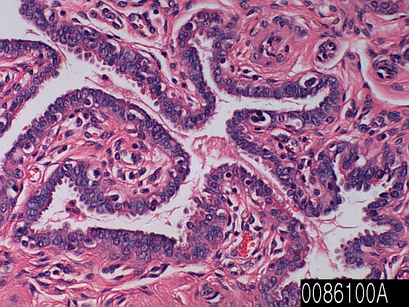
| Description | placenta, normal Age/Sex : 30/F |
| Price | 197 EUR |
| 260 USD | |
| 170 GBP |

Product Related Literature
Nutrition through the blood of the mother, waste removal, placenta is the organ that is associated with the development of the fetus to the uterine wall in order and to allow for gas exchange. , But also is a feature crucial for “mammalian placenta” or eutherian, placenta “true” has been found in lizards and snakes some with varying level of development to the level of the mammal. However, homology of structure, such as embryonic various organisms is questionable at best, in invertebrates such as arthropods such, best.The similar multiple, placenta is a classic no doubt, but the form placenta, has a wide currency at the moment and probably is common in modern English. In the language of the previous Rome tribal culture, to reflect the life mystery honored to be inherent in the birth process to bear fruit in the form of a child of cultural values often, placenta and “small” Mom “Grandma” or is called.
The mammalian Prototherial and (amateur) metatherial of (marsupial), while it is connected to the uterine wall, providing nutrients cocoon derived primarily produce placenta choriovitelline. Placental function as maternal fetal body that forms the placenta development mother (basal decidua) from uterine tissue of fetal and maternal, with placenta of fetal development (Shigeo chorion), the two components in the same blastocyst.
In humans, thickness from 2 to 2.5 cm of (0.8 to 1 in.) length and 22 cm placenta usually the center (9 inches), a thick, edge is thin. It usually weighs about 500 grams (1 lb). It has the colors of purple and dark red blue. It contains one umbilical vein and umbilical artery of two, and then connect to the fetus from the umbilical cord about 55-60 cm length (22-24 inches) it. (I have an eccentric attachment) to the umbilical cord insertion chorionic plate. Further separation to form a network that is coated on the surface vessels of the placenta branch, by a thin layer of cells. This results in the formation of villous tree structure. On the side of the mother, villous tree structure of these are grouped into lobules called cotyledons. In humans, the placenta is a disc-shaped common, but it varies greatly in mammalian species between different dimensions.
You can start developing blastocyst during implantation in the human endometrium in the placenta. Outer layer of the blastocyst becomes the trophoblast which forms the outer layer of the placenta. Syncytial the coating blastocyst and core layers: the outer layer is divided into layers of two additional. Are multinucleated cells a continuous layer covering the surface of the placental syncytiotrophoblast cells. This is a result of the ongoing process for synthesis of basal cell blastocyst differentiation and, in the development of the placenta. (Known as syncytia or otherwise) in the syncytiotrophoblast, and contribute to the barrier function of the placenta thereby. Placenta is increased through the pregnancy. Development of the blood supply of the mother, has completed by the end of the first trimester of pregnancy to placenta.
In preparation for implantation of the blastocyst, I receive a “decidualisation” endometrium. They become too complicated, its diameter is as though they were increased, spiral arteries in decidua has been remodeled. I act to increase the blood flow to the maternal placenta because straight flow with increased diameter. It can be a relatively high pressure, gas exchange maternal blood satisfies the villi space through the spiral arteries thereof is made, take a fetal chorionic blood. I in direct contact with the fetal chorion that other hemochorial human placenta and the maternal blood, the fluid is not replaced. Pressure, and pulse obezkisloroden between reduction of blood back through the veins of the endometrium.
IgG antibodies can be used to provide protection to the fetus in utero thereby pass through the human placenta. Begin as early as the 24th week of pregnancy 20th week, this transfer of antibody, of course. Look at the baby of the first few months of life of important ectopic as this, provides a toddler with a copy of the humoral immune long-term mother, this passive immunity, lasted several months after birth. The IgM antibody, however, infection of some acquired during pregnancy can not cross the placenta can be dangerous is the reason why the fetus. In addition, placental function as a selective maternal fetal transmission of the barrier to microorganisms. However, the failure of function, can cause the mother-to-child transmission of infectious diseases of mother still.