Seminal vesicle

Product name Seminal vesicle
Cat. No. 0077500A
No. of samples 1
Description seminal vesicle, normal
Age/Sex : 70/M
Price 197 EUR
260 USD
170 GBP

Seminal vesicle

Product Related Literature

Seminal vesicle small seminal vesicles (or gland vesiculosae) is a pair of simple tubular glands posteroinferior to the bladder of male mammals. They are located in the pelvis. The seminal vesicles, devote a large part of the fluid to be semen in the end. In lipofuscin granule secretion of epithelial cells dead, I get the color that the yellowish. It is not excluded in the fraction of the first zinc-rich prostate% of human semen derived from about 50-70 seminal vesicles is, and ejaculation, is dominated by the sperm. Once in the prostate, excretion tube of each seminal gland to open it in the appropriate vas deferens it. As a result of human sperm with a pH of weak alkalinity, semen is alkaline.

Alkalinity of semen helps to extend the life of the sperm, and to neutralize the acidity of the vaginal canal. I will be able to connect to the ejaculatory duct obstruction (pH <7.2) ejaculation acid solution. It generates a substance that causes the semen to become after ejaculation gelatinous / sticky, which is considered to be useful in maintaining close of sperm into the uterus to cells. Thick mucus seminal vesicles contain proteins, enzymes, fructose, mucus, ascorbic acid, flavin, a prostaglandin and phosphorylcholine. High concentration of fructose, which provides sperm of sustained energy during storage of sperm in the laboratory. It does not have to be likely to have contact with the semen vesicles screaming sperm in the vagina, but as the first step of their journey, they are transferred directly from the mucus of prostatic fluid through the female reproductive system. Liquid has been expelled the sympathetic contraction of the muscle layer.

Research in in vitro, that the sperm off pigs and fish eggs showed survival and motility poor and has been shown, it is protected low sperm chromatin. Therefore, the exact physiological significance of semen blisters unknown. If you want to form a low offset sperm plug the possibility of semen of men after arrival go to egg the last part of the semen, to guess as is the development of this recreation, yet, found in rodents some be

Covered with epithelium involved in the absorption and excretion (mucosa only mucosa or lining) or mucous membranes, it is lining, and endoderm origin mainly. They line the cavity is exposed to the external environment and internal organs. These are some places in contact with the skin: anal nose, mouth, lips, eyelids, ears, and genital area. The viscous secreted by glands called slime and mucous membranes, thick fluid. Term mucous membrane refers to where you are present in the fluid they were lining separation and body.

Clitoris hooded glans clitoris glans penis (penis head), is a mucous membrane and the inside of the foreskin. Is from the lining of the urethra. Preventing pathogens mucus trap body that is secreted, further act of the disease. It consists of longitudinal muscle layer of the inner and outer circular muscle layer in the layer outside. To prevent food from backward, longitudinal layer will reduce the tube of circular muscle layer. Rather than a layer of circular and longitudinal muscle true, layer, is stranded in a variety of fields. The inner circle is a spiral and outer longitudinal spiral and rugged terrain shallow pitch much.

Coordinated contraction of these layers, are called peristalsis move food through the digestive tract. Food in the gastrointestinal tract is referred to as (ball of food) bolus of the mouth into the stomach. After the stomach, was partially digested food, it is called chyme and viscosity. Colon semi-solid remaining is called feces.

It is Auerbach’s plexus or myenteric between two layers of muscle. This control peristalsis. Activity is initiated by pacemaker cells (interstitial cells of Cajal). Movement will have a (basic rate) intrinsic activity due to self-intestinal system. Rate, of course, can be modulated by other parts of the autonomic nervous system. Thickness of the muscle in the outdoors, differ in any part of the digestive tract. It requires a lot of power to feces is large, heavy, and press together, in the colon, for example, external muscles much thicker. The tapered vertical strips unrelated three outer longitudinal layer of the large intestine is known as (band of colon) plural form of F. taenia. This is one of the characteristics of three to help to distinguish between large and small intestine.

Weight loss segment of colon (2-3 times daily) You have to move all the way to the feces occasionally. If a person bowel movement remains, which is usually. Pyloric sphincter: pylorus of the stomach, is a thickening of the inner peripheral layer. In the gastrointestinal tract alone, and has an outer third layer of the stomach muscle layer. It is a layer of internal, this will help tilt the drum to the chyme in the stomach.

Prostate

Product name Prostate
Cat. No. 0077100B
No. of samples 1
Description Prostate, normal
Age/Sex : 54/M
Price 197 EUR
260 USD
170 GBP

Prostate

Product Related Literature

The Prostate is literally quite different among species anatomically, this is of most mammals It is a compound tubuloalveolar exocrine gland of the reproductive system of male, himself has been renamed the female prostate by the Federal International Commission on official autopsy term between the Skene’s gland or physiological 2002 female urethra near gland, and chemical.

It is said to be slightly larger than a walnut healthy human prostate classic. In the range of 7-16 g generally, average weight of the prostate “normal” in the adult male is about 11 ounces. It can surrounds the urethra below the bladder, just feel rectal examination. This is the only exocrine organ located in the midline in humans and other animals. Secretory epithelium is mainly pseudostratified cells, including basal cell and column, high is supported between the quality of elastic fibers, including the smooth muscle bundle orientation at random. Epithelium is highly variable, areas of squamous epithelium or low-cubic is present also, in the transitional epithelium in remote areas of the long tube. In the prostate, the supply called merged with prostatic urethra and ejaculation duct two urethra from the bladder. Prostate leaves or area, it can be divided in two ways. It does not have a capsule, but integral fibromuscular band surrounds it. These are coated with more pelvic floor muscles, ejaculation process.

Function of the prostate is to separate, slightly alkaline fluid that make up the 50 to 75% of the volume of semen in the liquid of the seminal vesicles and sperm normal, the appearance of white or milk. Semen, I made basic with total emissions from the contribution of other glands, including the seminal vesicle fluid of at least. The alkalinity of semen and helps to neutralize extend the life of the sperm, the acidity of the vaginal canal. Alkalinization of semen is achieved by secretion of seminal vesicles mainly. With the majority of sperm, prostatic fluid is discharged through the first ejaculation fraction. That compared to the sperm of a few that is discharged along with the semen blister fluid mainly, was expelled in prostatic fluid is, have a better protection of genetic material better mobility, and prolonged survival. The prostate contains the smooth muscle of some help expel semen during ejaculation.

Prostatic urethra is developing pelvic urogenital sinus of (endoderm origin) from (central) part. Endoderm outgrowths arise from prostatic urethra, growth in the surrounding mesenchyme. Glandular epithelium of the prostate, characterized by mesenchymal that is associated with the endoderm cells are characterized by smooth muscle and dense connective tissue in the prostate. Is a modification of the wall of the proximal part of the male urethra, prostate, derived from nine weeks of life of the embryo in the development of the reproductive system. Without mesenchyme, to fuse with the urethra, I condense the Wolf bile lead to adult prostate constitution body components closely glands and glands of some.

Prostate cancer is one of the most common cancers that affect the (estimated by some experts with 3%) the main cause of death in older men and older men in developed countries. However, “still, the potential benefits of the test, does not prove that exceed the harm of testing and treatment research” and, “They that person without learning the position of the American Cancer Society on the early detection I do not know about the benefits and potential risks of testing and treatment we believe that it should not be tested and know. (your prostate to 65 years before the brother and father of your race or black so that you can determine whether this is the right choice for you, for a discussion of the advantages and disadvantages of the test, talk test your doctor, and started 45 years) from 50 years old in the case you get a cancer. “If check is made, they may be in the form used to confirm the presence of (EN2) protein Engrailed-2 urinary measurement of prostate specific antigen physical rectal examination, the blood or (PSA), .

Richard Morgan and co-workers Hardev Pandha, announced his findings in the inspection of EN2 in 1 issue March 2011 urine of the journal Clinical Cancer Research. The clinical examination, to identify the current EN2 urine, home test kit was performed similar to home pregnancy test strip. According to Morgan, EN2 test is not yet available “We, and are preparing a large-scale study of some in the United States and the United Kingdom, but shows interest some companies to take it forward . “

Ureter

Product name Ureter
Cat. No. 0073000A
No. of samples 1
Description ureter, normal
Age/Sex : 40/M
Price 197 EUR
260 USD
170 GBP

Ureter

Product Related Literature

In human anatomy, generate smooth muscle, ureter is a tube made of urine from the kidney to the bladder. In adults, long (10-12) 25-30 cm usually, ureter 3-4 millimeters – diameter. The histological, further smooth muscle layer of support peristalsis third and remote transitional epithelium is included in the ureter. In humans, before you descend to the bladder in front of the psoas major muscle, ureter, coming out of the renal pelvis inner side of each kidney. Ureter, across the edge of the pelvis near the junction (they are passing priority) and the iliac artery. This is a common place for the congestion (the rest of the ureter bladder transitional valve meets the pelvouteric junction and bladder meet the ureter renal pelvis, the renal hilum to ureter) of kidney stones.

Run to the wall of the bladder for a few centimeters, the junction of the ureter and bladder, ureter executes the side wall of posteroinferiorly pelvic curve anteriormedially then to enter the bladder in the back,. Return of urine is prevented by valves known as the ureter bladder transitional valve. I receive the uterine artery and mesometrium through the bladder ureter in women. “Water under the (vas deferens or uterine artery) bridge (ureter) the phrase” effective to remember this anatomical relationship is. different channels play the same role in fish and amphibians, but the ureter was found in species of amniote all other. You can move from the kidney, and kidney stones, is introduced into the urethra, this has the potential to cut off the flow of urine, causing the lower abdomen severe cramps, or back side,. It is blocked you can develop a hydronephrosis of the kidneys, kidney affected, has become swollen due to the flow of urine.
Usually, hysterectomy is a surgical removal of the uterus performed by the gynecologist. (Called removal of the uterus body, and, “supracervical” also, leaving the cervix intact), hysterectomy (removal of the body, cervical and bottom, often referred to as “full”) general or it may be partially. This is the Department of Obstetrics and Gynecology of surgery most commonly performed. 90% is made under the condition of benign hysterectomy more than 600,000, were performed in the U.S. alone in 2003. These rates are the highest in developed countries has led to considerable controversy hysterectomy is unfair, that has been carried out mainly because of the unnecessary. If you become a can not (as in the removal of ovaries and fallopian tubes) patients bear a child, is not available treatment options other you have the long-term effects and risks of surgery, removal of the uterus, the surgery is typically recommended. In many cases, there is a better option, it is, that the incidence of hysterectomy is reduced for non-malignant indications are to be expected.

In many cases, (removal of the ovaries) ovariectomy, is achieved using a hysterectomy in order to reduce the risk of ovarian cancer. However, recent studies that prophylactic oophorectomy without emergency medical instructions to reduce the long-term survival of women have severe side effects, substantially free of the other has been shown [4], this effect It is not intended to be limited to premenopausal women. However, women who have already entered have demonstrated that it reduced the long-term survival in ovariectomized after menopause.

Hysterectomy may be performed in various ways. Technology known the oldest, is the incision in the abdomen. Then, technology laparoscopic vaginal later (do a hysterectomy to vaginal canal) vagina (instrument inserted through a small opening near the navel often, additional) have been developed. This is done by (not to be confused abdominal incision, and laparoscopy) abdominal hysterectomy most in the United States. Usually, near the upper limit of the hair in the lower pelvis pubic individual like incisions made for cesarean but as possible, (Pfannenstiel) incision is carried through on the stomach wall side.

It enables greater access to doctor reproductive structures, this technology was carried out in order to remove the whole reproductive complex usually. Because of the need to cut through the abdominal wall by sometimes more, recovery time for a hysterectomy the open, is 4-6 weeks. Historically, the biggest problem with this approach was infections, infection rate, has become a major problem in modern medical practice of which is not well controlled. Open hysterectomy provides the most effective way for you to explore the abdominal cavity, to perform complex operations. Before analyzing the laparoscopic vaginal techniques vaginal and it also, in most cases, vaginal technique is the only possibility to achieve desired contrast subtotal hysterectomy.

With the development of laparoscopic technology of 1970-1980 period compared to the procedures of the abdomen, surgery and minimally invasive has gained great popularity among gynecologists “laparoscopic vaginal hysterectomy” is (LAVH) after recovery is much faster. In addition, it allows a slightly more complex operation and inspection good procedure of the vagina. LAVH is finished begin with laparoscopy, (with or without removal of the ovaries) as is done through the vaginal canal removal of the ultimate of the uterus. Thus, LAVH a total hysterectomy, you may need to be removed in the uterus neck. “Laparoscopic hysterectomy with the supracervical” (Rush) of the cervix using a divider that can affect the uterus into small pieces that can be removed from the abdominal cavity through the post-laparoscopic port without having to remove and has been developed for the removal of the uterus.

Kidney

Product name Kidney
Cat. No. 0071000C
No. of samples 1
Description kidney, normal
Age/Sex : 43/M
Price 197 EUR
260 USD
170 GBP

Kidney

Product Related Literature

Kidney is an organ that acts as an important regulatory some invertebrates including some in vertebrates most. They are essential in the urinary system, maintain the regulation of the electrolyte such that the acid-base equilibrium plays a homeostatic functions (such as vias to maintain the water balance and salt) regulation of blood pressure. Providing a body as a natural filter of the blood, they removes waste that is diverted to the bladder. The flow of urine, were separated, the waste, such as ammonia and urea, they also kidney is responsible for the reabsorption of amino acid water, and glucose. The kidney is, I produce the hormone, including enzyme renin calcitriol, and erythropoietin,. The discharged renal vein that receives blood from the renal arteries located in the back of the abdominal cavity to the retroperitoneal, kidney paired, paired. The pairing structure in the sky urine, the bladder itself to each kidney ureter.

While renal physiology is a medical professional relationship, kidney, is the study of renal function in kidney disease. Kidney is different, but often, people with kidney disease shows clinical characteristic symptoms. Clinical common symptoms, including kidney, including nephrotic syndrome and nephritis, renal cysts, acute kidney injury, chronic kidney disease, urinary tract infection, kidney stones, urinary tract obstruction. Various types of kidney cancer exists, kidney cancer most adult is renal cell carcinoma. It can be controlled by removing renal disease other tumors, cysts and, a nephrectomy or kidney. Measuring renal function may be always a bad case, dialysis, choice of kidney transplantation therapy by glomerular filtration rate as a. They are very harmful, kidney stones, can be annoying and painful. Then, removal of kidney stones involves sonication to divide the rock into smaller portions that pass through the urinary tract. Common symptoms of kidney stones one is acute pain in the inside next segment of the waist.

In humans, in particular channel lie peritoneum and paraspinal, I was placed into the abdominal cavity is located at a position kidney was slightly tilted angle. 2, there is one on either side of the backbone. Usually, the asymmetry is slightly lower than the left in the abdominal cavity caused by the liver leads to the right kidney, left kidney is disposed slightly inside the right. T12 spinal cord, the left kidney is about slightly lower-right level to L3. Just in the right kidney at the bottom of the rear and rear to the left liver and diaphragm, the diaphragm under the spleen. This is the adrenal gland is placed on top of each kidney. Part upper part of the kidney (the skull) are protected by 12 ribs and 11 partially, adrenal gland and kidney of the whole, is surrounded by (fat pararenal and adrenal glands) layer of fat in two, renal fascia each have. 155 g and between 115 men and between 125, each adult kidney weighs 170 grams in women. Left kidney is slightly larger than the right kidney is typically.

It has the structure of kidney bean shape, and having each kidney uneven surface. Concave, renal hilum is the point at which renal artery enters the renal vein or organ, left ureter. Fibrous tissue severe kidney itself is surrounded by fat adrenal perirenal fat, renal fascia and (of Gerota), are surrounded by the renal capsule. Between forward muscle horizontal stripes are the boundary line of the rear (rear) and (front) border of these tissues is the peritoneum. Edge is adjacent to the liver and spleen to the left kidney on the right kidney. Therefore, I will move down the intake of both.

In the renal cortex on the surface, is a deep brain kidney: kidney or real, material is divided into major two structures. Obviously, such a structure, each contained around the renal cortex of the brain called the (Malpighian) renal pyramids may be in the form of renal sheet cone 8 to 18. There are projections of cortex called (of Bertin) renal pyramids between renal column. Functional structure of the medulla and renal cortex cover nephron, and urine production. Pass tubule, initial filtering of the nephron is the renal corpuscle position, and cerebral cortex followed from medulla pyramid deep cortex. Some of the renal cortex, medulla rays, is a collection of the renal tubular drain to collect channel.

To empty the urine into a small cup, teat or tip, of each pyramid, minor calyx of the sky to the major calyces empty renal pelvis, is in major renal calyx and ureter. Renal artery while entering, is in the gate, renal vein and ureter, end the kidney. Is a lymphoid tissue and lymph node hilar fat about the structure of these. Is located next to the cavity filled with fat, called renal sinus fat is hilar. Calices and renal pelvis are included together renal sinus, I have separated these structures from renal medullary tissue.

The kidneys receive blood from the renal artery, the left and right from the abdominal aorta branches directly. In spite of the relatively small size of their kidneys receive approximately 20% of cardiac output. Branch of the renal artery, which penetrate the renal capsule leading to areas artery is divided further into interlobar arteries extend through the columns of the kidney during the pyramid kidney. Interlobar arteries after the blood flow to the arterial arch that passes through the boundary of the medulla and cortex. Each arc-shaped artery is supplying interlobular artery and some of its feed supply afferent arterioles the glomerulus. Interstitial space is functional in the kidneys filter the individual is rich in blood vessels (glomeruli). interstitum to absorb the liquid, which was recovered from the urine. In various conditions, may lead to the failure or renal insufficiency and scarring, which can lead to congestion in this area. Take place in order to move through a small network of veins filtration of blood was collected in between leaf veins after. to return to the leaf between veins come to form the end renal vein the kidney for transfusion as well as the arterial veins follow the same pattern of interlobular, to provide blood to the arcuate veins then.

Rectum

Product name Rectum
Cat. No. 0068000A
No. of samples 1
Description rectum, normal
Age/Sex : 53/M
Price 197 EUR
260 USD
170 GBP

Rectum

Product Related Literature

The (meaning straight intestine from Latin rectum) rectum, in mammals, are linear and the last part of the colon in the gut of the other. Long about 12 centimeters (4.7 inches), (S-shaped end of the colon) rectum S-shaped junction, rectum of human begins at the level of the sacral promontory, sacral, by using the definition of third. Its diameter is the same as the S-shaped colon at the start, it has expanded to its end, to form the rectal ampoule. Depending on the definition that is used again, it ends at the level of the dentate line (or at the level of the puborectalis sling) anal ring. In humans, the anal canal is followed in front of the digestive tract rectum ends with anal verge.

The rectal intestinal act as a site for storing temporarily feces. It is placed in the rectum wall to stimulate the urge to have a bowel movement As the expansion for the material, so as to extend from receptors in the nervous system, rectal wall is full of inside. If it is not that desire to take action on the material in the rectum, in many cases, water is absorbed from the feces, to return to the large intestine. Constipation for a long time, defecation, if you delay the curing shit result. When the rectum is met, it is possible that the feces to increase the pressure to enter the groove, to force the rectal wall of the anal canal each other. Rectum force the anal canal is short as material, has been driving the feces from the rectum peristaltic wave. Sphincter of internal and external can be passed by muscles pulling the stool anus feces exit.

It is possible for the diagnosis of a particular disease, to perform a rectal examination. Suppositories may be used as a method of administering a drug, is inserted into the rectum. Endoscopes and colonoscopy S-shaped colonoscopy procedure is carried out for the diagnosis of diseases such as cancer. Insert rectal irritation fingers, was used to induce motion in a patient is sufficient to completely empty the peristaltic reflex rectal own rectum. Manual evacuation in the long-term management of neurogenic bowel seen most often in people who are using a gloved finger to evacuate the feces from the rectum, with multiple sclerosis and spinal cord injury and acute constipation It is used mainly.

(From the anus of the Latin word meaning is from, that proto Indian means “ring” “ring, anus” a) open the other end of the digestive tract of the mouth of the animal anus. Its function is, by desirably produced semi-solid, and controls the release of feces during digestion, and may contain, depending on the species: Inability to digest animal such as bone and nutrients to all food substances are extracted, for example, cellulose, lignin, it remains in the digestive tract, killed, or absorption problems that the toxic symbiotic otherwise intestine and more.

I use a mating and oviposition and (known as the cloaca) hole for separating the same solid waste and liquid amphibians, reptiles, and birds. I have a cloaca, which is considered Monotremates mammals also inherited functionality from amnion of therapsids via oldest. For separating solid and liquid, and marsupials, has an opening in the vagina particular for female reproductive. There is a hole that defecation, urination, completely independent for playback placental mammals women, channel flowing to the orifice has been almost completely separated, but the bowel movements of both the play and urination, men, another I have an opening.

Development of the anus was an important stage in the evolution of multicellular animals. It is likely that you followed a different path in the old and new mouth mouth animal, it would happen at least twice. Metamerism expertise is repeated later as head of the arthropod body of most bilateral body plan, and the body cavity, such as configured in fusion is built “module”,,, professionally: this is, promote or segment with the trend of significant evolution of the other.

In animals, the Haraguchi to deepen to become the first growth stage gastrulation, intestinal least, on the one hand, such as a complex, such as a worm embryos to form a recess. The new animal mouth, bowel tunnel option to be the anus for the opening of another in the mouth the initial dent. Because it was thought to be formed in the opening from the other end of the bowel later the anus and (“first” proto means) High hollow opening their first, it is called as the old mouth animal was. However, recent studies have shown that closed leaving an opening at their ends in the anus or mouth, an end portion of the center pilot opening old animals.

Colon

Product name Colon
Cat. No. 0067000A
No. of samples 1
Description colon, normal
Age/Sex : 60/M
Price 197 EUR
260 USD
170 GBP

Colon

Product Related Literature

The colon is the last part of the digestive system of vertebrates most. If you want to until it is eliminated from the body, to absorb water and salt from solid waste, they will occur where it is using the fermentation of unused material (mainly bacteria) plant. In contrast to the small intestine, colon, does not play an important role in the absorption of nutrients and food. However, large intestine does not absorb some fat-soluble vitamins water, and sodium. (Proximal colon refers to the transverse colon and ascending colon in general) S-shaped colon ascending colon, transverse colon, and descending colon: In mammals, the colon, has four sections. Anal canal colon, rectum and the cecum, colon before.
And behind it in the retroperitoneum or (intraperitoneal) and the location of the abdominal portion of the large intestine. They are to be secured to the village in general, retroperitoneal organ does not have full coverage of the peritoneum. Surrounded by the peritoneum completely organs in the abdominal cavity, and is therefore mobile. Rectal colon, ascending colon, and descending colon are intraperitoneal S-shaped colon cecum, appendix, and transverse colon, @ peritoneum. Because it affects the organs that can operations such as laparotomy such easy access to the inside, which is important. the colon of haustra colon, there is a small bag that is caused by vesicle formation give the appearance that segmented the. F. tapeworm runs the length of the colon. E. tapeworm because it is shorter than the small intestine, colon Nari has a tapeworm haustra formation between vesicles.

Arterial supply of the large intestine comes from the low branch mesenteric artery (IMA) superior mesenteric artery and (SMA). Flow between the two systems communicate via the “arterial limit” is parallel to the large intestine for its entire length. Able Historically, it is a variable vessel connecting the proximal SMA meandering mesenteric artery (of Witz), the proximal IMA arc or if Riolan that blocked the other hand, is that it is very important . However are considered, a recent study made in imaging technologies are improved, it has questioned the existence of the ship, some experts, for the removal of conditions from the medical literature future I’m called on. Affects the mesenteric vein over to connect the spleen to form veins lower hepatic portal vein poured into splenic vein usually arteries large and mesenteric veins, after venous drainage, liver it I go into. Lymphatic drainage of two thirds proximal rectum and the entire colon is, then the flow of para-aortic lymph nodes, the tank chyli. Rectum and anus of the other lymph is, you can either follow the same route, and to drain the superficial inguinal lymph nodes and internal iliac. Only rough, comb line marks this transition.

It is part of the four parts of the large intestine of the ascending colon. This first part of the colon a portion of the intestine such called the cecum, associated with the small intestine. Pass eight inches to about through the abdomen to transverse colon from the ascending colon. The primary function of one of the colon, is that to remove the other essential nutrients and water from the waste to be recycled back into the body. Said output of waste in the small intestine in the cecum, and then are migrated to the ascending colon, the recovery process will begin. Waste, is moved to the cross section of the large intestine by a process known as peristalsis.

It is from colon (large intestine from the end of the spleen) transverse colon, liver, spleen bending deflection. The connection will hang it by the broad band of tissue called the omentum transverse colon, stomach. And is attached to the rear portion of the mesentery of the abdominal wall is known as the transverse ligament on the rear side of the transverse colon. Coated in the peritoneum, therefore it, (not part of the colon just before and after) the transverse colon are mobile phones. To form the stool more generally, cancer forms, called (water removed) and colonic content becomes stronger. Two thirds close to the transverse colon, the third of the latter, is flooded downtown artery colic branch of the SMA while it is supplied by a branch of the IMA. “Turning point” between the blood supply of these two, is an area sensitive to ischemia represents the embryologic division between the hindgut and midgut.

there is a difference in the large intestine of organisms between different. The colon, it is possible to maintain the water balance, providing a space for the plants to absorb some vitamins such as vitamin C, using the fermentation, is responsible mainly for storage of wastewater reclamation. 90% of the water has reached the nutrients chyme, most at the time of this tube is absorbed by the body. At this stage, for example, sodium, magnesium, chloride and as part of the electrolyte (e.g. indigestible portion, amylose starch that is protected from the dietary fiber is significantly digestible carbohydrate digestion and the large left and eating food insoluble or soluble form or part). When the colon through chyme movement, most of the water remaining is removed, mixed with (known as intestinal flora) and slime bacteria porridge, it is converted to feces. Ascending colon preparation of fecal material in the form of a liquid. Then, move the muscles of the colon, forward aqueous waste, to absorb the excess water all gradually. Feces was solidified slowly to move along the descending colon them. In order to decompose a portion of the fibers to feed its own, and supplies in turn, as waste that is used in the lining cells of the colon, the bacteria will create propionic acid, butyric acid, acetic acid. No protein is not provided. In humans, in other animals, including primates and other monkeys with columns 10% larger undigested carbohydrates in proportion thereby probably become available, it is provided that allows a large part of the plant material in the manner more diet that is. Is an example of symbiosis, offers one day about a hundred calories for the body this [citation needed]. The colon and does not produce digestive enzymes – chemical digestion is completed in the small intestine before the chyme reaches the large intestine. PH in the colon varies between 7 and 5.5 (neutral weakly acidic).

Appendix

Product name Appendix
Cat. No. 0066000A
No. of samples 1
Description appendix, normal
Age/Sex : 46/F
Price 197 EUR
260 USD
170 GBP

Appendix

Product Related Literature

The application is a blind tube type that is connected to the cecum since been developed embryology. It is pouchlike structure cecum, the colon. Annex is located near the junction of the small and large intestines. Term “appendix” means “form of worms” and comes from the Latin. It has evolved independently marsupial diprotodont, in monotremes, and also widely present in Euarchontoglires, size and shape is very diverse it. The length of the human appendix is ​​AVG 11 cm, but may be in the range of 2-20 cm. The diameter of the application is a 7 ~ 8mm typically. I was measured 26 cm from the patient Zagreb, Croatia further deletion up until now. Application is located in the lower right quadrant of the abdomen close to the bone of the right hip joint. Position in his abdomen, which corresponds to a point on the surface, which is known as (see below) McBurney point. Core applications are cm ileocecal valve 2 following a fixed location relative to, the position of the tip of the application peritoneal (behind the cecum) cecal Yes (74%), may vary to be in the pelvis is out.

Description had been missing for lack of importance and unique shape of the appendix as determined by the long side does not exist, it is gut-associated, called the immune system surrounding the intestine and other places administrative The lymphoid tissue, has been accepted that you perform many important functions, but the effect of appendicitis after. Flash disease, bacteria of those Duke University, which was proposed by William Parker, Randy Bollinger, in 2007, from the rest of the bowel, which functions as a haven for bacteria application from colleagues. Immune system, including its architecture, in combination with the popular features of many applications, only this proposal, the growth of beneficial intestinal bacteria the position of the flow below the normal one-way bacteria and food of the large intestine association with copious amounts of immune system and is based on a new understanding of how to promote. In a study conducted at the University Hospital Winthrop, possibility that individuals that have not been added have a recurrence of four times C. difficile showed high. However, other studies, infection among people who have added those which do not indicate that there is a large proportion of C. difficile.

Possible scenario for the transition to the appendix of the human from the current function cecum completely around by Charles Darwin. Be used for administration to digest leaves as primates was suggested him. It may be evidence of ancient human anything has deteriorated almost in the course of evolution. As long cecum herbivores like some found in the koala and horse like this, it seems to confirm this theory. Cecum of the koala, will be able to host the bacteria to help it to decompose the cellulose specifically. The ancestors of the human race, you may want to rely on this system when I was living in green diet is rich in them. As people have started eating the food digestion easier, you may want to become less dependent on plant cellulose content of energy is high they. As was the need much to digest cecum, mutation as survived, mutation (interfere with the progress and evolution) is harmful before it does not matter. It that these alleles become more frequent, cecum has been shrinking was suggested. The cecum and the need, in millions of years later, this to degrade the applications of today.

The most common disorder of the application, I have a (appendix carcinoid) carcinoid tumor and appendicitis (of people). About 1 200, application cancer is a malignant tumor of the gastrointestinal all. In rare cases, I also present adenoma. Is a state (epityphlitis) or appendicitis is characterized by inflammation of the appendix. In response to the development of the embryo midgut pain and appendix of often, it starts in the center of the abdomen. Usually, dull, this pain is visceral pain that is poorly localized. Because inflammation progresses, pain will start localization more clearly inflammation and peritoneal quadrant, lower right. Peritonitis and inflammation of the peritoneum, leading to (pain upon removal of pressure than the application of pressure rather) recovery response. In particular, it presents a third of the way along a line drawn from the anterior superior iliac spine McBurney point, the navel. In general, pain points (skin) is not only displayed on the parietal peritoneum and is inflamed. Immune system and fever is typical of appendicitis.

Appendicitis will require removal by laparoscopic or abdominal inflammation or addition. If untreated, the application potential rupture shot followed and left untreated, death still lead to peritonitis. Surgical removal of the appendix applications are called appendectomy or appendix. If the patient is suffering from acute appendicitis, usually, this removal is performed as an emergency treatment. When the surgical facility does not exist, intravenous antibiotics are used to prevent or delay the onset of sepsis. More often, inflammatory mass formation around the appendix, appendicitis resolves completely in some cases. This is relatively contraindicated for surgery.

Recently, to deepen the understanding of gut immunity, refers to the ability of apparent mammalian cecum use, such as a safe-house for the plant protection on infection of gastrointestinal in society symbiotic gut, no modern medicine, cognitive biology I merged with and medical basis. Potentially, this feature is a selection force for the development and maintenance of applications. Be described in mammals based on the shape of the cecum mainly can be three-form type of cecum application: (as of primate species many) cecum, the upper part of the vast cecum, appendix as an appendix to the long , and application (dunes of Cape Moruratto and large wing rabbit in the absence of a clear cecum) and (wombat as) it is an important branch of larvae or round. Further, the elongated addition, or found mammals (like monotremes) is shown in or lacks the cecum, such structures are important between structures, such as applications and cecum (like Koala) junction Na is missing. The appendix cecum, it developed independently at least twice and is another example of the convergence in the form of placentals between marsupials Australia in the rest of the world is displayed. Application, but also have been lost by host species obviously being maintained for at least one strain 80000000 or more years.

Stomach antrum

Product name Stomach antrum
Cat. No. 0063000C
No. of samples 1
Description Stomach, antrum, normal
Age/Sex : 51/M
Price 197 EUR
260 USD
170 GBP

Stomach antrum

Product Related Literature

The cardia of the anatomical term of adhesion stomach, in the esophagus. Starting from the distal to the Z-line of the gastroesophageal junction as squamous epithelium of the esophagus in order to give way to the columnar epithelium of the digestive tract immediately cardia. (GE) junction is ambiguous anatomically close to the cardia of the stomach, but it is proven physiological lower esophageal sphincter. Called the lower esophageal sphincter overlap cardia, no cardiac lower esophageal sphincter does not include area. Topic, but tried to consensus of the members of the current is in the cardia of the stomach before. Overlap with the cardia, but does not include (also called esophageal sphincter sphincter, stomach and esophageal sphincter [5] heart) is the lower esophageal sphincter (LES), for example. In contrast and it is configured in the control striated muscles innervated by the body Accordingly, “upper esophagus sphincter” contained in the hypopharynx, and (extending from the base of the tongue zone cricoid cartilage). By being in the LES, Z lines, can be identified from the color of the Z-line of dark red from pink to change the LES. However, mirror localization endoscopic (Z line) was found to be 3 cm distal to the Z-line position of functional LES investigation and correlation internal pressure of LES.

And prioritization epitympanic middle ear cavity, the cone part of the temporal bone through the aditus to communicate (tympanic cavity mastoideum, cavity Valsalva) is back to the cells of the mastoid cavity in milk (milk input cavity) mastoid cavity the airspace. Air these spaces will provide the voice of resonance, functions as a sound receptor, functions as a sound diffusion, to provide protection from mechanical damage and reduce the weight of the skull. Mastoid air cell system, has contributed significantly to the inflammatory disease of the middle ear.

(Cavity, a small cul-de-sac) pyloric cavity is the first part of the pyloric region of the stomach. It lies at the bottom of the stomach near the pyloric sphincter to separate the stomach and duodenum. Exclusion from the rest of the stomach during digestion of about muscle Batch peristaltic contraction prepyloric fully or partially temporarily, this is the second of the pylorus stomach (pyloric passage) through the channel of some it You may from part, it is sometimes, it is defined.

Stomach produces strong acid and (hydrochloric acid) enzymes (such as pepsin) to help digestion. The digestion mixture are called gastric juice. Rather than the influence of the stomach itself, the inner lining of the stomach has several mechanisms to resist the lining of the esophagus. Normally, the esophagus are protected from these acids and the junction with the one-way valve mechanism stomach. The one-way valve, called (ES) esophageal sphincter, which is prevented gastric formed together from flowing back into the esophagus herein the angle of Cu. During peristalsis, ES bolus of food to be able to pass through the stomach. I will prevent to be returned mixture of digestive enzymes chyme, bolus, stomach acid and, through the esophagus it. It is assisted in its task of maintaining the material flow in one direction of the diaphragm to the ES. In ES, is the anatomy sphincter function but sphincter. Means that the thickening of the smooth muscle does not exist at all as pyloric sphincter such This is prevented travelleling back from gastric chyme the esophagus (usually). It helps to lower the CRU, to the diaphragm, the action of this sphincter.

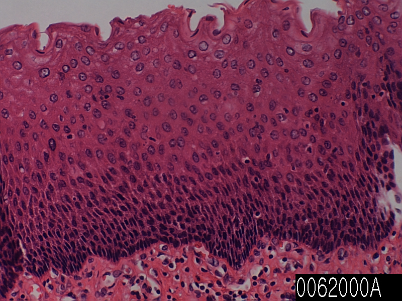
Esophagus

Product name Esophagus
Cat. No. 0062000A
No. of samples 1
Description esophagus, normal
Age/Sex : 33/F
Price 197 EUR
260 USD
170 GBP

Esophagus

Product Related Literature

Consists of a tube of muscle that passes from the stomach to the throat of food (known esophagus, as esophagus), esophagus is the organ of vertebrates. Peristalsis of stomach via swallowing, while traveling by moving from the mouth through the pharynx to the esophagus food. Words that are lit is derived from the Greek word derived from Latin oisophagos esophagus esophagus. In humans the “entrance. to eat”, esophagus is continuous with the level of the larynx pharynx C6 vertebrae. Esophagus passes through mediastinal back to the chest, abdomen entering through the opening of the diaphragm at the level of the tenth thoracic (T10). This is a 25 cm Normally, instability, recorded from 10 to 50cm, depending on the individual level. It is divided into the abdominal portion of the cervix, and breast. Because of the inferior pharyngeal constrictor muscle, it enters the esophagus open only to vomiting or when swallowing.

It is called the sphincter heart cardias sometimes or esophagus, but really it is not considered to be a valve, in fact, the junction between the (node ​​GE or gastroesophageal junction) stomach and esophagus, and more I’m similar to a good structure. The esophagus, in order to obtain a peristaltic wave that causes (called a bolus) balls food digestive tract of a sequence, the majority of contracts smooth muscle. In humans, it is a peristaltic contraction of smooth muscles to drive the contents through the digestive tract.

In most fish, the esophagus, very low mainly due to the (associated with the gills) the length of the throat. Not lungfish and lamprey chimera, true stomach, esophagus, so that it flows directly from the pharynx to the gut very effectively, and a little longer, however, some species,. To be, in short esophagus much longer than that of the fish according to the pharynx of tetrapods. In amphibians, in addition to the peristaltic movement of muscle, sharks and rays of the esophageal epithelium eyelashes, to help wash food together. Kang, esophagus is the poultry only in vertebrates most, to form a culture for preservation of food before entering the stomach True, it has been extended to the bottom. Structure with the same name connects the mouth to the stomach often, it is found in vertebrates, including arthropods and mollusks.

Is a thin layer of smooth muscle in the majority (muscularis mucosa) or muscularis mucosa [formula? In] gastrointestinal tract, is located on the outside of the lamina propria, and is separated from the submucosa. Refers to the muscularis mucosa (if it happens) the combination of the lamina propria and epithelium in the term “mucous membrane” or gastrointestinal mucosa. Etymology “is a special layer of the lining of their own,” as translated into Latin name I suggest “muscle layer of mucous membrane” and (lamina propria) (laminate muscularis mucosa), that. Muscle mucosa some of oriented smooth muscle fibers in a variety of ways to keep the surface of the associated glands and mucous membranes in a constant state of shaking slightly to expel the contents of the contact increased between the cavity contents and epithelium and crypt It consists of a thin layer.

(For example, respiratory tract, genitourinary tract and gastrointestinal tract) lamina propria is a component of the wet pad known as the mucosa or mucous membrane tube of various lines in the body. Lamina propria (ie, lamina propria mucosa) is located under the epithelium, which is a thin layer of loose connective tissue is mucosa and epithelium together. The name of the Latin shows, it shows that it is a characteristic component of the special lining of the lining ‘own coat. “Therefore, the. Specific layer relates to the combination of epithelial-specific plus laminate, contains the central lymphoid tissue in the small intestine and milk and capillaries (lymphatic),. Lining or mucosa term mucus secretion in the lamina propria always gland and duct to open lining epithelium of serous secretion and is included. lamina propria is rich in immune cells known as B lymphocytes. of IgA, the majority of these cells, secretion is a B-cell.

Human normal tissue

Cat. No.Product DescriptionNo. of Samples
0001000CSkin, breast, normal1
0004000CBreast, normal1
0004100ABreast, nipple, normal1
0007000ASpleen, normal1
0008000BLymph node, normal1
0013000ASkeletal muscle, normal1
0026000ABronchus, normal1
0028000BLung, normal1
0055100AParotid gland, normal1
0056000CLiver, normal1
0057000AGallbladder, normal1
0059000BPancreas, normal1
0061100B
Tonsil, normal1
0062000AEsophagus1
0063000AStomach fundus1
0063000CStomach antrum1
0064300ADuodenum1
0065100B
Jejunum1
0066000AAppendix1
0067000AColon1
0068000ARectum1
0071000CKidney1
0073000AUreter1
0077100BProstate1
0077500ASeminal vesicle1
0079200AVas Deferens1
0083000AUterine cervix1
0085000AMyometrium1
0086100ASalpinx1
0087000AOvary1
0088100BPlacenta1
0088800AUmbilical cord1
0093000AAdrenal gland1
0096000AThyroid1
0098000AThymus1