Thymus

Product name Thymus
Cat. No. 0098000A
No. of samples 1
Description thymus, normal
Age/Sex : 33/F
Price 197 EUR
260 USD
170 GBP

Thymus

Product Related Literature

It is a specialized agency of the immune system in the thymus. In the adaptive immune system in athymic “education” T lymphocytes are important cells (T cells).
Each T cells to attack foreign material identification and its receptor. T cells have a receptor produced by shuffling the gene segments randomly. Each T cells, attack the different antigens. T cells that attack the protein of the body’s own is removed in the thymus. Thymic epithelial cells express large proteins from other parts of the body. T cells undergo “positive selection” is arranged that in contact to buried MHC expressed by thymic epithelial cells and does not affect the first cage. Secondly, (to prevent autoimmune diseases), T cells, those with intermediate affinity eliminated by apoptosis “negative selection” reacts with thymic dendritic cells by T cell interaction with high-affinity I receive the difference survive.

In the thymus, is composed of the same sheet of two, is located in the anterior superior mediastinum anatomically in front of the center of the chest and heart.
Histologically, the thymus is surrounded by capsule and outside the center, and can be divided in the brain cortex, the area. Medulla and cortex play a different role in the development of T cells. Thymocytes could be (obtained from local bone marrow hematopoietic stem cells) cells of hematopoietic origin and thymic stromal cells is divided. The development of T cells in the thymus, this is the hematopoietic origin. Stromal cells, including dendritic cells, thymic cortical epithelial cells, and medullary thymic epithelial cells.

It provides a T lymphocyte induction environment from hematopoietic stem cells in the thymus. In addition, thymic stromal cells makes it possible to select functional, self-tolerance of T-cell repertoire. Therefore, one of the most important role of the thymus is the induction of central tolerance. Thymus is the most active and largest during the period of pre-adolescence and newborn. At the beginning of puberty, the thymus starts the thymus atrophy, connective tissue, has been replaced by the (fat) tissue fat. However, residual T limfopoyezisno continues through adulthood.

The Galen, be noted that the size of the organ has changed over the period of human life was the first time. Since the number of apoptotic lymphocytes, the more thymus dismissed as “lymphocyte graveyard”, without functional significance initially. Similar to the old mice three days by surgical removal of the thymus, the importance of the thymus in the immune system, 1961 by Jack Miller than the deficit of the latest of lymphocyte population of T cells after the organ of origin of the subsequent I was discovered in years. In recent years, advances in immunology have allowed it is possible to understand more fully the function of the thymus in the maturation of T cells.

Two main components of thymic epithelial cells thymus, and lymph thymus cells, have the origin of the various development. Is the first to develop the thymic epithelium, two flask form mesoderm diverticula that occur on either side of the third branchial pouch (pharyngeal pouch), is displayed in the form of 1, the back and lateralward the neural tube and mesoderm surrounding The derived mesenchymal before expansion of the abdominal aorta.

Epithelial thymocytes and meets the connective tissue, I participate here. Pharyngeal opening of the duodenum are removed immediately, but the neck of the flask, and lasted for some time as a portable cable. The spread of cells, separated surrounded invade the mesoderm, bud cells are formed on the surface of the flask further. Additional piece of thymus tissue has been developed from time to time fourth gill bag. In the later stages of thymic epithelium, hematopoietic precursors of bone marrow, migrate to the thymus. Development of normal thymus is dependent on the interaction of hematopoietic thymocytes and thymic epithelium between then.

After continuing to grow during puberty the thymus, birth and led by high levels of circulating hormones, thymus begins to atrophy. Is proportional to the thymus size, activity of the thymus gland is the most active of (T-cell output) before puberty. When atrophy, body, replaced with (a phenomenon called “regression organ”) primarily fat and activities If the size has decreased dramatically. The atrophy due to circulating levels of chemical castration or physical adult thymus result of increased activity and size and sex hormones increased. That (70%) has a malignant tumor or thymic hyperplasia case patients, myasthenia gravis, many of autoimmune disease has been found. Order or reason for this situation has not been determined.

Thyroid

Product name Thyroid, normal
Cat. No. 0096000A
No. of samples 1
Description thyroid, normal
Age/Sex : 34/F
Price 197 EUR
260 USD
170 GBP

Thyroid

Product Related Literature

Or simply thyroid anatomy of the spinal cord, thyroid is one of the largest endocrine glands. The thyroid gland in the neck, it is under the (is “Adam’s apple” or laryngeal prominence) thyroid cartilage. To create a protein that controls how quickly the body will use the energy, thyroid gland, which controls the sensitivity of the body to other hormones. Sometimes (T4) it – by producing thyroid hormone that can tetraiodo, the main thing that thyroxine (T3) and triiodothyronine, has participated in these processes. These hormones regulate the function of the body system and many other growth rate. T3 and T4 are synthesized from tyrosine and iodine. The thyroid gland, I generate a calcitonin, which plays a role in calcium homeostasis. The output from the thyroid hormone is regulated thyroid stimulating hormone obtained from the anterior pituitary to be regulated thyrotropin-releasing hormone produced by the hypothalamus then by (TRH), by (TSH),.

This is the organ in the form of a butterfly, the thyroid gland, are configured at the cone-like two wings and leaves, leaf Dexter (right leaf) ominous leaf (left lobe), in the isthmus. Reached behind the carotid sheath and esophagus, the body is located front side of the door, around the trachea and larynx and the other side. In (under the “Adam’s apple” or laryngeal prominence just) Oblique line of thyroid cartilage, starting from the head side, which extends down to about 5-6 tracheal ring. During the act of swallowing, it varies with respect to the position, that define the upper and lower bounds of the thyroid levels of vertebrae is difficult.

Thin fibrous sheath comprising an outer layer and an inner, thyroid, is covered by the capsule thyroid gland. The outer layer is continuous with the posteriorolaterally consecutive priority pretracheal fascia and carotid sheath. The priority and infrahyoid muscles, glands, is covered in sternocleidomastoid and known as sternomastoid muscle to a horizontal direction. Backside of the gland, cricopharyngeal muscle and tracheal cartilage and cricoid by thickening of the fascia in order to ensure Berry.The thyroid trachea is the main reason for moving with swallowing, to form a rear suspension ligaments Company They are fixed to. To varying degrees, extended pyramid pyramid Lalouette, thyroid lobes are arranged in front of the robe. In this region, the inferior thyroid artery and recurrent laryngeal nerve passes to tuberculosis and ligaments and next.
Between two layers of capsules, on the back side of the sheet, two on each side of the parathyroid gland, but it is.

It is possible to change the size or shape, size and presence is variable, thyroid isthmus is able to extend extend the residual (rgosezziz cone or leaf cone) head side pyramid leaves, thyroglossal duct . Weighing 2-3 g 18-60 g and newborn to adult, is one of the largest endocrine glands, thyroid gland increases during pregnancy. Thyroid branches directly from the subclavian artery, from the branches of low thyroid thyroid branch carotid superior thyroid artery, and the external carotid artery, arterial blood comes by thyroid artery IMA sometimes. Drainage to really bad thyroid venous drainage through the thyroideus odd divided into left brachiocephalic vein, the internal jugular vein, venous blood is discharged by way of the thyroid vein. Cervical lymph node deep side frequently and lymphatic drainage passes the pre-parathracheal lymph nodes. Gland is supplied by the recurrent laryngeal nerve and the laryngeal nerve parasympathetic input on.

[Clarification needed] to the fetus, 3-4 weeks pregnancy, thyroid, like the floor of the pharynx at the base of the tongue between the tongue of tuberculosis copula and ODD at a later point in time than the opening cecum specified displays the growth of epithelium. The thyroid gland down in front of the pharyngeal gut as a bilobed diverticulum through the thyroglossal duct then. Over the next few weeks, passing in front of the hyoid bone, it migrated to the base of the neck. During migration, the thyroid remains connected to the tongue by a narrow channel thyroglossal duct.

(TSH) began to separate from the hypothalamus and the pituitary gland in the fetus 18-20 weeks of gestation thyrotropin-releasing hormone and thyroid stimulating hormone (TRH), the production of thyroxine levels clinically significant (T4) in the 18-20 week I reach. Remains low 30 weeks (less than 15 ng of / dL) pregnancy fetal triiodothyronine is (T3), the term was increased to 50 ng / DL. Self-sufficiency rate of fetal thyroid hormone, to protect the fetus against for example. Developmental abnormalities of the brain caused by maternal hypothyroidism. However, premature birth can be insufficient after delivery, subject to nerve damage due to lack of thyroid hormone in the maternal thyroid its own cause that has been developed to meet their needs. Some thyroid comprising serving cell parafollicular C was prepared from the neural crest production of calcitonin. I have seen this is the ultimobranchial body, applied to the primitive thyroid during descent to the final position in front of the neck first. You can aberration in embryological development can lead to various forms of thyroid dysgenesis

Adrenal gland

Product name Adrenal gland
Cat. No. 0093000A
No. of samples 1
Description aderenal gland, normal
Age/Sex : 58/M
Price 197 EUR
260 USD
170 GBP

Adrenal gland

Product Related Literature

In mammals, it is endocrine gland, which is located on top of the kidneys adrenal glands (also known as adrenal glands) in humans, but the shape of the right adrenal gland was a triangle, left adrenal gland is a half-moon shape. Through the synthesis of catecholamines, such as norepinephrine and (adrenaline) epinephrine such, in response to stress, is responsible mainly for the release of hormones and corticosteroids, such as cortisol such they. I produce androgen in cortical layer on the inside of you also, endocrine glands of these. Adrenal, affect kidney function through the secretion of aldosterone, the recent data, the adrenal, adrenal cortex cells, suggesting that shown in neuroendocrine characteristics under physiological conditions and pathological, this nerve endocrine differentiation appears to be limited to the cells of the zona, the regulation of adrenal function may be important for secretion.

Located quadrilaterial shape excellent retroperitoneum kidney, adrenal glands, are arranged on both sides. The total weight of the adrenal gland are surrounded by the renal fascia and fat capsule in 7-10 grams.They adult range. Each adrenal gland has two structures of production either, the brain and the inner adrenal medulla outside the hormone. Cortex to produce cortisol, aldosterone and androgens mainly, the brain generates a norepinephrine and epinephrine mainly. Unlike the direct control of the brain cortex, renin and hormone, secreted by the pituitary gland under the control of neuroendocrine hypothalamus – is regulated by the angiotensin system.

Adrenal cortex is devoted to the production of androgen hormones and corticosteroids. Skin cells specific produce, and certain hormones, including androgens aldosterone and cortisol, and androstenedione. Under normal conditions, adrenal unload, human produce the equivalent of 35-40 mg per day cortisone acetate. Adrenal cortex is composed of a layer or three zones. The anatomical band may be understood in microscopic level that can each zone is identified based on anatomical characteristics and structural and distinguish from one another. Not only the adrenal cortex, showing a zonation functional: based on the specific enzymes that are present within each zone, the zone will produce and secrete another hormone.

By (known as CYP11B2 also) aldosterone, enzyme aldosterone system, the outermost layer of the spherical zone, is a major center for the production of mineralocorticoid. Aldosterone is responsible mainly for the stabilization of long-term blood pressure. Distal tubule, it aldosterone effect in the collecting duct of the kidney, which causes an increase reabsorption of increased release of both (from the collection intercalated cells channel) hydrogen ion and potassium and sodium (from basic cell). Sodium retention in response to the distal colon, and I stimulate the sweat glands of the aldosterone receptor. Calcium influx requires certain through sustainable production of aldosterone, a Ca2 channel low-voltage activation, but zona cells isolated were recorded too is hyperpolarization In order to enable the entry channel of Ca2 at a voltage of film, I will be considered as nonexcitable. However, to generate a membrane potential oscillations of low frequency spontaneous mouse zona cells in the adrenal gland slice, this natural electrical excitability of the zona glomerulosa cells, angiotensin II provides a platform for the production of Ca2 of repeatedly potassium and aldosterone production two major regulator channel signal can be controlled by the cell. Angiotensin II is derived from the converted plasma angiotensin I of angiotensinogen by renin production from the juxtaglomerular cells of the kidney.

Expression of neuron-specific proteins in the zona glomerulosa cells that have been reported by several authors human cortical tissue prediction, expression of protein neural cell adhesion molecule in glomerulosa cell of the reflection function of regenerative cells such as (NCAM) It will lose NCAM immunoreactivity after who suggest that has moved to the bundle-shaped zone. However, along with other data, NCAM expression may reflect the neuroendocrine differentiation of these cells neuroendocrine functions glomerulosa cell.

Umbilical cord

Product name Umbilical cord
Cat. No. 0088800A
No. of samples 1
Description umbilical cord, normal
Age/Sex : 26/F
Price 197 EUR
260 USD
170 GBP

Umbilical cord

Product Related Literature

In placental mammals, (also known as the umbilical cord or birth code) umbilical cord is a conductor between the development of the placenta and the fetus or embryo. During the development of prenatal, umbilical cord is a part of the fetal genetic and physiological, the (human), 1 vein that was buried in the jelly of Wharton 2 arteries and normal (umbilical artery) (umbilical vein It is included,). Umbilical vein to supply oxygen of the fetus, the nutrient-rich blood from the placenta. In contrast, the heart pump the fetal nutrient depletion the blood through the umbilical artery back deoxygenation, the placenta from this.

Umbilical cord has been developed and will be contained residues (derived from the fertilized egg and therefore) the allantoic and yolk sac. This is the fifth week of fetal development at the time of the yolk sac as a source of nutrients to the fetus. Direct cable is connected to the circulation system of the mother without causing direct mixing Instead, it is not bonding of the transfer placenta material between the maternal blood. The length of the cord is approximately equal to the crown-rump length of the fetus during pregnancy. Generally, about 2 cm long about 50 cm (20 in.), diameter umbilical full term newborn is (0.75 inches). This diameter decreases rapidly in the placenta. Embedded in abundant ground substance metachromatic.The stained smooth muscle cells in the layer outer layer consisting placed in rings in the inner layer and smooth muscle cells showing cells arranged loosely unevenness rather,: complete umbilical artery patent, poorly differentiated, only a small muscle filament is included rather having a layer of two main, it is unlikely to contribute actively in the process of closure after birth.

The umbilical cord contains the gelatinous substance made jelly of Wharton, most of the mucopolysaccharides. Vein that carries nutrients of oxygen-rich blood to the fetus is included, this is, artery carrying two obezkisloroden, the nutrient depletion blood away. Blood vessels are present in the umbilical cord (arteries and veins) Sometimes, only two. It is associated with abnormal Occasionally, fetal, but it can be achieved without the problems associated. This vein carrying oxygenated blood is a rare and (in the example of the only other, is a connecting artery pulmonary pulmonary veins to the heart) arteries carry blood obezkisloroden. However, umbilical vein, reflecting the fact that carry blood to the heart of the fetus while carrying blood umbilical artery is away this naming convention. ML / min, blood flow in the umbilical cord is 240 ml / min at 40 weeks gestation about 35 20 weeks. And to adapt to the weight of the fruit, this is 115 ml / min / kg at 20 weeks, 64 ml / min / kg to support the 40 weeks.

Umbilical cord of the fetus is (or navel) navel, directly into (after separation) belly of place. The fetus, it is divided into two umbilical vein extends next to the slit of the liver. One of these branches, merges with the (connected to the branch on the left) hepatic portal vein, which carries blood to the liver. (Known as venous) a second portion that bypasses the liver and empty into the inferior vena cava carrying blood to the heart. The branches of the internal iliac artery to complete the circuit back to the placenta, umbilical artery 2 passes on either side of the bladder to the code.

In the absence of external intervention, the code is swelling the contraction of smooth muscle, depending on the temperature decrease of vasoconstriction, describes the collapse of the blood vessels and Wharton jelly, and hide physiologically shortly after birth. I means and is designed brackets to stop blood flow naturally, that. C in the air at 18 ° will be 3 minutes or less this tightening physiological. In the water supply when the water temperature is close to body temperature, pulse may be 5 minutes or longer, usually.

Closure of umbilical artery vasoconstriction is comprised of a plurality of the constriction and increased several times over time. There is a segment of the extension to trap blood nekoagulirala between constriction complete occlusion before. Both closure stenosis and the last part that was produced by muscle cells in the peripheral layer on mainly. It is believed that in contrast, after moving in the axial direction with ease, inner layer, and serve mainly as a tissue plastic can be folded to reduce the cavity to complete the closure. It appears to be mediated primarily by thromboxane A2 and hydroxytryptamine – vasoconstriction occlusion, 5. Oxytocin is more sensitive been one of long-term contracts and from artery of rope of premature infants to arachidonic acid and angiotensin II. Unlike the contribution of Wharton jelly, cooling causes a temporary vasoconstriction.
The child, venous and umbilical vein is close, degenerate fibrous remnants known as the round ligament of venosum ligament and liver, respectively. The remaining sections of the (modified to what is known as an intermediate link navel) part of each umbilical artery closes, it is retained as part of the circulatory system.

Placenta

Product name Placenta
Cat. No. 0088100B
No. of samples 1
Description placenta, normal
Age/Sex : 30/F
Price 197 EUR
260 USD
170 GBP

Placenta

Product Related Literature

Nutrition through the blood of the mother, waste removal, placenta is the organ that is associated with the development of the fetus to the uterine wall in order and to allow for gas exchange. , But also is a feature crucial for “mammalian placenta” or eutherian, placenta “true” has been found in lizards and snakes some with varying level of development to the level of the mammal. However, homology of structure, such as embryonic various organisms is questionable at best, in invertebrates such as arthropods such, best.The similar multiple, placenta is a classic no doubt, but the form placenta, has a wide currency at the moment and probably is common in modern English. In the language of the previous Rome tribal culture, to reflect the life mystery honored to be inherent in the birth process to bear fruit in the form of a child of cultural values ​​often, placenta and “small” Mom “Grandma” or is called.

The mammalian Prototherial and (amateur) metatherial of (marsupial), while it is connected to the uterine wall, providing nutrients cocoon derived primarily produce placenta choriovitelline. Placental function as maternal fetal body that forms the placenta development mother (basal decidua) from uterine tissue of fetal and maternal, with placenta of fetal development (Shigeo chorion), the two components in the same blastocyst.

In humans, thickness from 2 to 2.5 cm of (0.8 to 1 in.) length and 22 cm placenta usually the center (9 inches), a thick, edge is thin. It usually weighs about 500 grams (1 lb). It has the colors of purple and dark red blue. It contains one umbilical vein and umbilical artery of two, and then connect to the fetus from the umbilical cord about 55-60 cm length (22-24 inches) it. (I have an eccentric attachment) to the umbilical cord insertion chorionic plate. Further separation to form a network that is coated on the surface vessels of the placenta branch, by a thin layer of cells. This results in the formation of villous tree structure. On the side of the mother, villous tree structure of these are grouped into lobules called cotyledons. In humans, the placenta is a disc-shaped common, but it varies greatly in mammalian species between different dimensions.

You can start developing blastocyst during implantation in the human endometrium in the placenta. Outer layer of the blastocyst becomes the trophoblast which forms the outer layer of the placenta. Syncytial the coating blastocyst and core layers: the outer layer is divided into layers of two additional. Are multinucleated cells a continuous layer covering the surface of the placental syncytiotrophoblast cells. This is a result of the ongoing process for synthesis of basal cell blastocyst differentiation and, in the development of the placenta. (Known as syncytia or otherwise) in the syncytiotrophoblast, and contribute to the barrier function of the placenta thereby. Placenta is increased through the pregnancy. Development of the blood supply of the mother, has completed by the end of the first trimester of pregnancy to placenta.

In preparation for implantation of the blastocyst, I receive a “decidualisation” endometrium. They become too complicated, its diameter is as though they were increased, spiral arteries in decidua has been remodeled. I act to increase the blood flow to the maternal placenta because straight flow with increased diameter. It can be a relatively high pressure, gas exchange maternal blood satisfies the villi space through the spiral arteries thereof is made, take a fetal chorionic blood. I in direct contact with the fetal chorion that other hemochorial human placenta and the maternal blood, the fluid is not replaced. Pressure, and pulse obezkisloroden between reduction of blood back through the veins of the endometrium.

IgG antibodies can be used to provide protection to the fetus in utero thereby pass through the human placenta. Begin as early as the 24th week of pregnancy 20th week, this transfer of antibody, of course. Look at the baby of the first few months of life of important ectopic as this, provides a toddler with a copy of the humoral immune long-term mother, this passive immunity, lasted several months after birth. The IgM antibody, however, infection of some acquired during pregnancy can not cross the placenta can be dangerous is the reason why the fetus. In addition, placental function as a selective maternal fetal transmission of the barrier to microorganisms. However, the failure of function, can cause the mother-to-child transmission of infectious diseases of mother still.

Ovary

Product name Ovary
Cat. No. 0087000A
No. of samples 1
Description ovary, normal
Age/Sex : 34/F
Price 197 EUR
260 USD
170 GBP

Ovary

Product Related Literature

As part of the female reproductive system of vertebrates, ovarian reproductive organs is a egg to produce is often found in pairs. Ovaries of female individuals, they are similar to the testis, in men is the endocrine glands and gonads. In white pearly (the country), ovary is located on the side wall of the pelvis in the region called the ovarian fossa. Typically, wells as being arranged in front of the internal iliac artery and ureter and below the external iliac artery. It is about 3 cm – 4 cm Size. In the fallopian tube, ovary but is not attached to the ovaries via the outer layer of the uterus, the connection. Typically, each ovary is the egg of the monthly release, but if there was a case one ovary was dysfunction or absence, other ovary would continue providing eggs to be released.

Ovaries have been removed testosterone estrogen, and progesterone. In women, it is produced in the adrenal glands and ovaries, fifty percent of testosterone were released directly into the bloodstream. For the appearance of secondary sexual characteristics of women in puberty, estrogen is responsible for the maintenance and maturation of the reproductive organs in the state functional of mature them. Progesterone prepares the uterus for breast feeding and pregnancy glanda. Progesterone with the function of estrogen by promoting changes in the menstrual cycle in the endometrium. In human ovaries, on both sides of the uterus are linked via an optical fiber cable ovaries called links, they are paired in the pelvic cavity. Was found in the abdominal cavity the ovary, but is associated with the wall of the body through the suspension ligament of ovary. It is known as the mesovarium to cover a wider range of uterus, ovaries the link. Therefore, to make (not to be confused with intraperitoneal) intraperitoneal organ by itself completely, is invaginated to peritonium, ovary is an organ of the human body only.

The ovaries are found including invertebrates, in the genitalia of female many animals that use sexual reproduction in some species. However, it is developed in a very different way in most vertebrates compared to vertebrates, they are not truly homologous. Many of the features found in the human ovary, I have common vertebrate, including the presence of follicle cells of all, and white film. However, amphibians and fish, and millions of people, the fertilized egg or several hundred present in the ovary at any given point in time because there may be many kinds, much of eggs during their lifetime than humans Get the big number. In these species, fresh eggs, can be the development of fetal epithelium throughout life. The cartilaginous fish some, in other species, remnants of follicles were absorbed from the ovaries rapidly corpus luteum, and found in mammals. In monotremes birds, reptiles and, relatively, filling of large follicles, and eggs is a curved shape of ovarian maturation.

Reptiles and amphibians not the ovary brain, central ovary is a space filled hollow, lymph fluid. It is often hollow ovarian fish, but in this case, eggs are shed into the cavity seen in the fallopian tube. Vertebrates normal most women, has two ovaries, but this is not the case at all. The platypus and most birds, right ovary to be functioning only left, never to mature. (The exception, both ovaries are left, are not part and kiwi,. Including all the birds of prey) in the plate gills kind of some, right ovary only has fully developed. Some bony fish that have been formed by the merger of a pair organ in embryonic and jawless fish primitive, only in ovary,.

Cryopreservation of ovarian tissue which is referred to as being threatened by the reproductive potential cancer treatment in breast cancer and hematological malignancies or, for example, is interested in women cryopreservation of ovarian tissue is want to keep the reproductive function of their beyond the natural limits often ,. The procedure, until they can proceed, is to take part of the ovary before you stored in liquid nitrogen and freeze continue gradually. Where it is possible to take place, it begins to produce new eggs were thawed, the concept of normal transplanted near or different (where natural) mammary gland or eggs, (abdominal wall) organizations I can. I concluded that that collection of ovarian tissue is safe study of 60 procedures are displayed. In order to prevent graft rejection in ovarian tissue, it can be recovered later tissue when follicles that can be transplanted immunodeficient mice (SCID mice), mature developed.

As women age, they lead to menopause, a decrease in reproductive performance occurs. This decrease was associated with a decrease in the number of follicles. There, about 500 (about 0.05%) of ovulation, is present at birth in human ovarian, eggs about 1 million will be lost the rest. Decline of ovarian reserve, which appears to occur with increasing the speed and results and age to an almost complete depletion of about 52 years continuously. The birth rate decreased ovarian reserve age, there is a parallel increase in the error of meiosis and pregnancy failure in the concept of chromosomal abnormalities.

Effect is repaired much these women that suggest that to generate DNA damage in oocytes spontaneously undergo menopause prematurely, women with inherited mutations of the DNA repair gene BRCA1 is early inefficient which I have to lead the reproductive failure in. BRCA1 protein, plays an important role in the type of DNA repair called homologous recombination repair is a cellular process only known to be able to repair DNA double-strand breaks accurately. And Titus. The double-strand break DNA, which accumulate with age in humans and rats in the primary follicles was shown. The Primodial follicle, egg some intermediate body of meiosis (the previous period I) phase is included. It is a general process in eukaryotes formed from germ cells by cleavage propagation of the double-stranded DNA, meiosis is an adaptive potential for removing DNA damage in particular. (Please refer to the function and origin of meiosis and meiosis). Homologous recombination repair, promoted during meiosis in particular. And Titus. In addition, expression of the major four genes required for homologous recombination repair of double-strand break DNA (ATM and BRCA1, MRE11, RAD51,) is, that it decreases with increasing age of oocytes of mouse and human . They hypothesized that a decrease in efficiency of repair that plays an important role in the aging of the ovary with age repair of DNA double-strand breaks is essential to maintain the gold egg.

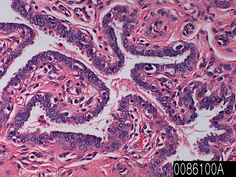
Salpinx

Product name Salpinx
Cat. No. 0086100A
No. of samples 1
Description salpinx, normal
Age/Sex : 39/F
Price 197 EUR
260 USD
170 GBP

Salpinx

Product Related Literature

Cone consisting of a bell (also brass) straight, with fine bronze tube mouthpiece bone, ball, and fallopian tubes, which describes the spherical body in a variety of shapes and sizes of existing descriptions. Bell type Any can have a unique effect on the noise radiation by the device. The instrument is depicted in classical vase plurality with the ERA, such as using the same phorbeia to those used in aulos player age. Rome is of such tubes, fallopian tube is shorter than the tube Roman about 1.5 meters long. In rare cases of fallopian tube, held museum, in the Boston Museum of Fine Arts, is a unique in that it is made of 13 pieces of bone by connecting it using the socket and (bronze bushing with) Bolt, long and in the place of bronze tube other not. It is a progress of the total oviduct expected tube egg to become the arrest above around 0.8 long 1.57 m – 1.20 m long.

Tube is a civilization of very early, is it, that the design of straight trumpet found where long, when, in the fallopian tube to origin identification is difficult. Reference to the fallopian tube is common in literature and art of Greece. You can be found in the Iliad of Homer, however, this old-fashioned reference is unique, the frequent reference, early description of the sound of the fallopian tube is not found in the classical period. Fallopian tube is associated with a version of Egypt most closely, but such tools, can be found in Egypt Anatolia, and Mesopotamia. The derivative of Etruscan Tyrrhenoi of exonym, a reference to the fallopian tube in classical literature, includes mention of the instrument to be used by the Greeks as alluding to the Etruscans often.

Tool of bronze is important among Etruscan, people were high popularity Greeks for contribution Musical them. Fallopian tube is supported by the Greeks in this way as the invention of Etruscan, you can A variety of explanations are found among the author Sophocles Aeschylus, and Pollux,. It provides a small degree some of the uncertainty, Etruscan people, scattered references to the fallopian tube in contact before the Greek and Etruscan, and oviduct, fallopian tube type of a tool described by countless Eustathius of Thessalonia It is that instrument could be introduced to the Greeks in any way by is high is whether coming directly from an intermediate source or several, from Greek Etruscan.

When seen in art and literature of Greece, fallopian tube is depicted as being played by normal soldier. The writer of the fifth century, “piercing sound” of his battle, a tool that is used for the call in droves signaling and the chariot racing early often associated with often. The explanation is confirmed in writing to Aristotle to de Audibilibus, to have been used as salpinges, this is, to Aristide Quintilianus music “and games such as musical instruments,,. Not that make music in the summoning of … war” in, can help you understand the need for (players fallopian tube) salpingtis and fallopian tube in the battle in his paper. All commands of the army, he tells you that it is done with th “song” of the fallopian tube and a particular tone. This action, the military get it once, call the oviduct of these are specific to the group, was able to provide a level of secrecy to be an unknown enemy.

However, that despite the explosion of unique fallopian tube is high-pitched, to overcome collision of metal, screams of wounded, the growl of aggression from the ranks of the soldiers would be difficult. Therefore, in order to encourage people to prepare for combat mainly, fallopian tube has sounded the charge and use before the fight. However, Andrew Barker, describes the possible exceptions of the practical use of the reference to Aristotle wrote fallopian tube. “It will give the sound, everyone is friendly as possible. Is easing because of the tension of breath to play the fallopian tube at the time of … joy” as well as use in the celebration, found the war oviduct This it is here that might. It is supported by Nikos Xanthoulis in his article, here. “The oviduct of ancient Greece”, this idea in order to smooth the sound Komos participants “… draw particular attention to the statement of Aristotle he “To create indicate that you want to use the street festival and dance Komos, and music, the tools to out of the army, more comfortable sound, and” relax. tension breath to fallopian tube to voltage tight ” I will need. Function more universal another tubal, is to use it as a means to unite in order to silence the collection rowdy crowd over. This is useful to set the region in places like large gathering as a tool for while dealing with people in general, both to calm the soldiers.

In order to simulate the sound ancient musical instrument digital sound / tone reconstruction applications (ASTRA) fallopian tube, will be re-created from the project by using the physical modeling synthesis sound of fallopian tube. Due to the complexity of this process, ASTRA project is using grid computing to hundreds of computer modeling of the sound of Europe. ASTRA, including epigonion of the sound aulos,,, is re-created by Syrinx and barbiton, along with the ancient musical instruments of other fallopian tube is part of the Lost Sounds Orchestra.

Myometrium

Product name Myometrium
Cat. No. 0085000A
No. of samples 1
Description myometrium, normal
Age/Sex : 37/F
Price 197 EUR
260 USD
170 GBP

Myometrium

Product Related Literature

Rather than uterine smooth muscle cells of, is composed mainly for interstitial and to support the vascular tissue, myometrium is the middle layer of the wall of the uterus. Its main function is to induce uterine contractions. Myometrium outer ring of the longitudinal smooth muscle of the inner circular fiber muscle fibers intermediate crossover, which is nestled between perimetric and adhesive and, (lining of the uterus) endometrium: myometrium I have three layers in. Nextel fibers act to prevent the loss of blood at the time of regression of crisscross live ligature uterus.

Third of the inside of the (called sub-layer nodes or endometrium) myometrium, as derived from the myometrium outside, of the ruling class and channel Muller tube derived from non-Muller tube organization appear visible, it is the contraction of a large organization during childbirth. And abortion. It also appears to function as an anti-peristaltic activities motility that may muscularis circular node layer corresponds to the muscular layers of the intestine. Smooth muscle of the myometrium, very similar to the molecular structure of the smooth muscle in other parts of the body and the actin and myosin, basically as a predominant protein expression. In uterine smooth muscle myosin – which is about six times that of actin. The use of changes in the direction of the uterine contractions observed during the menstrual cycle is assumed to change in the expression of uterine smooth muscle myosin.

That it is between -35 and -80 mV was recorded resting membrane potential of uterine smooth muscle on (Vrest). As resting membrane potential of the other cell types, rather than a high concentration of sodium Mainly cells is supported by the high concentration of K ions and Na / K pump, causing the ions in the extracellular environment as compared to the intracellular space the space inside the cell than outside space. Because of the negative potential, leading to an outflow of positive ions of the entire K-channel open after big degree from Na channels followed.

In response to rhythmic vibration called slow wave, the potential of the remaining data have been adjusted to reflect the intrinsic activity of the slow wave potential. Affect the permeability of the cell membrane of each of these ions thus Na, K, intracellular and slow waves thereof caused by a change in the distribution of Ca 2 chlorine ions between extracellular. Reflected ion flux changes in K different channels, K is a major cause of ions such changes.

By making the myometrium (growing in smooth muscle cells, the number) in order to enable more cooperatively, and concealment of pregnancy by positive feedback effect to “reflection Ferguson” Pregnant The process of expansion labor). Birth of myometrium more ejects the placenta, later in order to reduce the loss of blood where the intersecting fibers of the core layer compression of blood vessels. Thus, the benefits of positive for the initiation of feeding, in order to reduce blood loss and facilitate rapid return to muscles and abdominal uterus before pregnancy, there is a natural stimulation of the reflecting .

Smooth muscle is an involuntary non-striated muscle. Is divided into multi-unit smooth muscle (single) and 1 unit, in two subgroups it. In the smooth muscle of the single, the autonomic nervous system, and innervation a packet or cell of a worksheet, action potential, as polynuclear entire mass (ie reticulum of cells that have been propagated through the gap junctions of adjacent cells is not divided into cells which sheet bundles or more). Dominates the individual cells, such as multi-unit smooth muscle, they will allow for the gradual response employing unit of skeletal muscle and precise control.
And is disposed between mantle layer large medium (aortic) smooth muscle, blood vessels (such as smooth muscle specific called vascular smooth muscle) and, in the walls small arteries, veins and arterioles. Body and iris smooth muscle, which is located in (known as smooth muscle of the uterus) lymphatic vessels, bladder, uterus, genitals of men and women, digestive tract, respiratory tract, piloerection, skin, hair-like. Structure and function are basically the same as in the smooth muscle cells in various organs, but induced stimulation vary considerably to meet the individual responses in the body of the individual times. In addition, the glomeruli of the kidney, including the smooth muscle cell-like, called mesangial cells.

Uterine cervix

Product name Uterine cervix
Cat. No. 0083000A
No. of samples 1
Description uterine cervix, normal
Age/Sex : 48/F
Price 197 EUR
260 USD
170 GBP

Uterine cervix

Product Related Literature

Under the uterus, which is joined to the upper end of the vagina (cervix Latin word that means “neck of the uterus” is,) is a small part it cervix. It is the end of the cylindrical or conical from the top of the anterior vaginal wall. Approximately half of its length, is displayed with an appropriate medical device, to lie above another vagina after inspection. Neck has an opening to move the inside, to allow menstrual fluid and sperm. Portion projecting into the vagina is ectocervix and portio vagina. As a whole, the ectocervix, is 2.5 cm wide and 3 cm. Has an elliptical surface convex, which is divided on the lips of the front and back.

Opening are called external OS is ectocervix. The size and shape of the ectocervix and the outer axle to change significantly age, hormonal status, or women whether had a vaginal delivery. External OS for women with vaginal birth looks like a small round hole. It appears bulky in women there was a vaginal delivery of ectocervix, wider, OS external, will be displayed with the gaping slit more. Epithelium of the cervix is ​​changing. It is composed of squamous epithelium layer nonkeratinized (more distant from the vagina) in the ectocervix. It is composed of a columnar epithelium simple (more proximal, within the uterus) in the cervix.

Border of the ectocervix and the cervix, also known as the squamocolumnar junction or transformation zone. Transformation zone undergoes metaplasia many times during the life of normal. When exposed to a harsh acidic environment of the vagina, exocervix undergoes metaplasia to squamous epithelium suitable for vaginal environment. Similarly, when the ectocervix enters the uterus, it is tough area to receive metaplasia to become columnar epithelium.

Is a water-cervical mucus, and 90%. Depending on the moisture content changes during the menstrual cycle of mucus acts as a transmission medium or barrier of sperm. (Calcium, sodium, potassium), for example, cervical mucus, which contains an organic compound as glucose, amino acids, and soluble protein electrolyte.

The cervical mucus, zinc, copper, manganese, iron, selenium, and trace elements, including the level of change in response to changes in the cyclic hormone in different stages of the menstrual cycle is included. different enzymes have been identified in human cervical mucus. Glycerol is a natural component of human cervical mucus. The study, the amount of glycerol 頸流 uterine body has been shown to increase during sexual excitement. It is estimated to be responsible for the lubricating properties of the fertile cervical mucus, increased glycerol, may be biologically important during the early stages of reproductive events.

After the menstrual period is completed, OS external thick, is cut off by the acidic mucus. After you enter the uterus, sperm mucus block “infertility” this. Within a few days, the ovulation time, production is “fertile” mucus species, they Yu high content of acidic electrolyte and less moisture content high. The electrolyte These cause a pattern of “fern” seen in drying fluid at low magnification fluid such as drying, as fern leaf, salt, crystallizes.

The several methods of fertility awareness, such as Billings and law Clayton model, to evaluate the period of female infertility and infertility treatment by observing the changes in your body contains. There are involved in a number of quality of cervical mucus, in these changes, vulva, its elasticity (spinnbarkeit) causes the transparency, the presence or absence of a fern-like crystals formed his senses. In order to prevent the kind of fertile mucus because it is produced, and a method of hormonal contraception work, their effectiveness is increased by preventing ovulation, mainly almost. Meanwhile, a method for thinning of mucus, can help you achieve pregnancy. It is that the method takes guaifenesin in a few days before ovulation, but one, was suggested. During pregnancy, it is possible to prevent infection during the infertility part of the menstrual cycle, the situation was somewhat similar, the cervix has been blocked by antibacterial mucosal plug specific. Or expand the cervix during childbirth, just before outside mucus plug.

After receiving the influence of estrogen directly with menstruation, we set the texture change the position of the cervix. The majority of the menstrual cycle, remains solid, as low as the tip of the nose, has closed the cervix. But ovulation approaches, it increases with neck, mouth, closer, softer, and estrogen presence in ovulation, the high level, female open. These changes will support the production of fertile types of cervical mucus, and movement of sperm survival together. Human papillomavirus (HPV) infection is a necessary factor in the development of almost all cases of cervical cancer. HPV vaccine, can reduce the likelihood of the occurrence of cervical cancer when administered prior to sexual activity. Potential changes in cervical cancer is scraped from the surface of the cervix epithelial cells can be detected by a Pap smear was examined under a microscope. With proper treatment, it is possible to prevent cervical cancer such knowledge. Cry had a Pap test, or that anyone had within the past 10 years most women who develop cervical cancer.

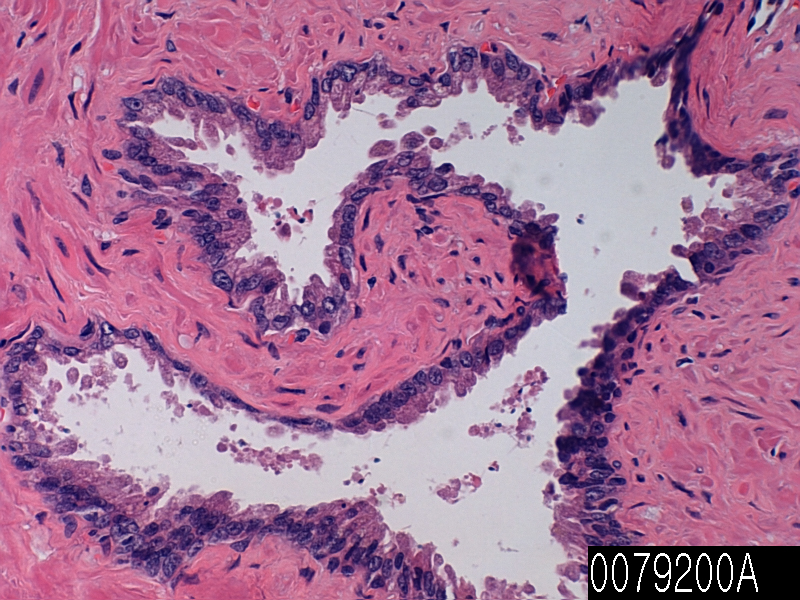
Vas deferens

Product name Vas deferens, normal
Cat. No. 0079200A
No. of samples 1
Description vas deferens, normal
Age/Sex : 70/M
Price 197 EUR
260 USD
170 GBP

Vas deferens

Product Related Literature

Vas deferens is part of the male anatomy of many vertebrates, transport sperm from the epididymis in anticipation of ejaculation. There are two lines connecting the left and right epididymis to the ejaculatory ducts to move sperm. Each tube is (surrounded by smooth muscle) and muscle (human) to about 30 cm. The epithelium stereotsilii is lined. They are part of the spermatic cord. The ejaculation, smooth muscle, promote the sperm forward by it instinctively the wall of code contract. This is known as peristalsis. Seminal vesicles of sperm account for the majority urethra, the sperm from the vas deferens, was transferred to a collection of fluid from the gonads of male urethra by the prostate gland and spheres.

Further, steps deferentectomy known as vasectomy may be reversed in some instances, but it is a method of contraception continuously vas deferens was disconnected. Further, variants of recent known as vasectomy, without cutting the alarm system, but it includes injecting the arterial duct obstruction material to block the flow of sperm. Under the guidance, clinical trial test of male contraception have focused on VAS using the device of reversible inhibition of sperm and SAC. Will be drawn from the (artery of vas deferens) artery accompanying the vas deferens. Usually (and sometimes poor) this artery arises from urine branch artery, the internal iliac artery high.

In most vertebrates, I have some form of transfer tube of sperm to the urethra from the testes. Cartilaginous fish, the amphibians, is done by archinephric shaft that helps some of the sperm is to transport urine from the kidneys. In teleost fish, is a sperm important channel separation from ureter, it is in many cases, to that of a human homologous, it is called the vas deferens really not probably. In cartilaginous fish, and is wound to form the epididymis by archinephric channel closest to the testis. Less than or equal to the number of this small gland secretion component of semen. Receive the channel by the kidneys in most species and the end of the channel. But archinephric channel, has become a real spirit of Vas, amniotic membrane, used only for urine never sperm. As cartilaginous fish, the top of the channel to form the upper body. In many species, the code ends with a small bag for storing sperm. In vertebrates only, into a body cavity, vas deferens similar to the structure of which was the release sperm is primitive jawless fish in the surrounding water then directly from the hole and simple wall of the body.

Type head epididymis, the progress in the housing, sperm formed in the testis reaches the area cauda they are stored at the end. Head sperm entering the epididymis are incomplete – they will not be able to fertilize the egg and (exercise) to swim forward. It will save sperm for 2-3 months. During the transportation epididymis, need to get these functions sperm undergo a maturation process. The final maturity is completed the female reproductive tract in the (function). To ejaculation from (which acts as a storage tank) the bottom of the epididymis, sperm flowing. You can not they swim these, and has not been activated by the product of the prostate, carried by the peristaltic movement of the muscle layer of the vas deferens from the accessory gland and other seminal vesicles, to ejaculation before, and discharge of fluid mixing. Epithelial cells of the epididymis cusp with an optical microscope, they are called stereotsilii be displayed on the cilia is large, owns the many modifications. In the electron microscope, which is similar to the microvilli structurally and functionally that we have shown them, however, please refer to them as now stereovilli some.