Prostate adenocarcinoma Gleason’s Score 9

Product name Prostate adenocarcinoma
Cat. No. 7781403A
No. of samples 1
Description adenocarcinoma
Age/Sex : 59/M
Price 197 EUR
260 USD
170 GBP

Prostate adenocarcinoma Gleason's Score 9

Product Related Literature

Gleason grading system was to evaluate the prognosis of men with prostate cancer. Incorporated strategies prostate cancer stage helps to predict the other parameters, the estimated values ​​together, to guide the treatment. Gleason score is given to the prostate based on its microscopic appearance. Cancer is more aggressive and high Gleason score, it is a poor prognosis. In most cases, radiologists and urologists, through the rectum using a biomedical scientist in the laboratory histology prepared microscope slides for immunohistochemistry for diagnosis by pathologists and H & E and needle, a hollow I remove a cylindrical sample of prostate tissue and (biopsy). Prostate, after being removed during surgery, a pathologist will slice the prostate for inspection.

The pathologist is assigned a Gleason score based on the sum of the two numbers. It is a rank of tumor models the most common, is the second number, the first number is the degree of the most common model of second. If the model of three of the first number is present, the most common and, second, is the highest class. For example, tumor model is the most common third degree, but it will be some cells is a fourth grader has been found, Gleason score 3 + 4 = 7. The second number, which is a slight change in the Gleason system pre-2005 is the level of the secondary cell lines most common. This class also known as Gleason pattern, or Gleason score, and is known as a sum Gleason. With the prognosis of five worst Gleason pattern and Gleason grade in the range of 1 to 5. Gleason score is in the range of 2 to 10, 10 to the worst prognosis. Cancer more aggressive Gleason 4 +3 and evaluation of Gleason 7 3 +4 Gleason. In addition, there is no really difference between the tumor or 10 attack assessment Gleason 9.

Then, in order to obtain a final result, these materials are added grease. Increasingly, pathologist, to provide evidence of “high” component. Where just a few of the 1 (Normal attack) model of 3 minutes. Therefore, there may be 3 +4 tertiary Gleason parts of the scheme to make it 5 – would be considered it, and prostate cancer more aggressive in Gleason 3 +4 5 without high-end models. Although it is a question as a certain degree of any full component highly malignant cancer. I have never seen almost 2 in the form of current, system Gleason, and prostate cancer Gleason pattern 1, and Is the rarely seen and live by definition. Gleason pattern 3 is the most common by far. So 3 +3 = 6 Gleason score, it is considered as a class of cancer can be seen in practice, usually, these cancers should have a pretty good estimate actually.

The scoring system, named pathologist in the development Minneapolis Veterans Affairs hospital is equipped with colleagues at other facilities in this Donaldson, in 1960. In 2005 from the international community of urinary pathology, Gleason, change the system. The sophisticated, standards, has changed the decision of some models. Incidentally, has a higher performance compared to the original Gleason score modified “above, it standard that is acceptable to the urological pathology current has been demonstrated. In this form, it remains important tools some.

Prostate cancer is a form of glandular cancer onset, of the male reproductive system in the prostate. Growth is slow prostate most cancers, but in some cases of invasive cancer of the prostate. Tumor cells can be from prostate other body parts, especially in lymph nodes and bone to (spread) transition. Prostate cancer, the problem may be pain, difficulty urinating, during intercourse, or cause erectile dysfunction. Other symptoms, may occur during the later stages of the disease potentially.

Is detected less frequently than South Asia, East Asia, in Europe, in the United States, detection rate of prostate cancer varies greatly throughout the world especially. Prostate cancer tends to develop in men over the age of 50. Globally, (currently, the first of the second in the United Kingdom and the United States) it is a major cause of the sixth of cancer-related death in men. Prostate cancer is most common in developed countries has increased in developing countries. However, many men, not treated without symptoms never, prostate cancer die because of other unrelated eventually. Many factors, including the Food and genetic is involved in the development of prostate cancer.

It can be shown by biopsy presence of prostate cancer, symptoms, physical examination, prostate specific antigen or (PSA),. The prostate-specific antigen test, detection of cancer will increase, but it does not reduce the death rate. Because of the risk of retreatment and overdiagnosis, such as prostate cancer, U.S. Preventive Services Task Force will remain asymptomatic, which is recommended for screening for prostate cancer using the test PSA in 2012. USPSTF concluded that the potential benefits of the test is not exceeded expectations harm.

Management strategy for prostate cancer, should be guided by the severity of the disease. I can be low-risk tumor to track active surveillance very safely. For curative treatment, typically do not include surgery according to radiotherapy and various normal or cases of advanced disease rarely more, cryosurgery, hormone therapy, chemotherapy, and the (hormonal therapy Regardless, in some cases, it is reserved for applying a radiation of) possible. Several studies, masturbation, has been shown to reduce the risk of prostate cancer.

About human age, and health, of metastasis, appearance, and the base of the response of the cancer to initial treatment are important in determining the prognosis of the disease under the microscope. Decision of whether to treat (cancer contained in the prostate) localized prostate cancer patients and radical is a compromise between the adverse effects to the expected beneficial to survival and quality of life of patients.

Usually do not cause symptoms early prostate cancer. But sometimes, the symptoms are similar to these diseases as benign prostatic hyperplasia such frequent prostate cancer. These include (painful urination) dysuria to start the steady stream (nocturia) frequent urination frequent urination at night, difficulty urine, maintain, and, hematuria (blood in the urine) is included . About one-third of patients diagnosed with prostate cancer between asymptomatic, two-thirds have more than one of these symptoms.

Prostate cancer is associated with dysuria prostate that surrounds the prostatic urethra. Therefore, changes in the gland can affect the urinary function directly. Secretion of the prostate and seminal of the urethra vas deferens deposits prostate as included in the content semen, prostate cancer, it’s performance sexual function and as difficult to obtain ejaculation and painful erection of such can cause problems.

Advanced prostate cancer, which can cause additional symptoms probably spread to other parts of the body. In many cases, (bones of the spine) spine, ribs and pelvis, the most common symptom is bone pain. Toward the proximal portion of the bone, cancer, spread to the bone of others, such as the femur in general. Prostate cancer of the spine, can be pressure and weakness of the legs, leading to fecal incontinence and urine, and spinal cord.

Bladder papillary transitional cell carcinoma

Product name Bladder papillary transitional
Cat. No. 7481303A
No. of samples 1
Description bladder, papillary transitional
Age/Sex : 62/M
Price 197 EUR
260 USD
170 GBP

Bladder papillary transitional cell carcinoma

Product Related Literature

Papilloma grows (projection) exophytically in fingerprint list refers to a benign epithelial tumors. In this context, instead of the projections created by papillomas, pointing (for example, cereals) papillary tumors, existing. When used without connection to this, in many cases, it is those infections caused by (HPV) warts, human papilloma virus related (squamous cell carcinoma),. However, other conditions that cause papilloma, there is not a known cause, and the number of cases a lot of it. The HPV infection in most cases, there is no cancer that occurs, but human papilloma virus infection is a major cause of cervical cancer. Papillary epithelial origin benign tumor. Prediction arising from the mucosal surfaces of the cauliflower-like. It may be normally white or colored. This may be fixed or pedicle. The average size is less than 2.0 cm. I do not like strong for sex. The most common site in the palate, lips and tongue followed by uvula area. Period of up to 10 years a few weeks.

Renal papilla is a urine anywhere in the marrow pyramid sky in a small cup in the kidney. And wherein the convergence marrow manifold pipe so as to form a flow path Bellini to direct fluid histologically. To start as can be seen in transitional epithelium. chemicals or toxic to some of the kidney called nephrotoxins is, exercise their renal papilla. Renal papillae can lead to the death of cells in this region of the kidney called the renal papillary necrosis. The most common cause of toxic renal papillary necrosis is a non-steroidal anti-inflammatory drugs such as ibuprofen combination, aspirin, phenylbutazone, and dehydration. Pancreatic duct and common bile duct is perforated the middle portion of the second portion of the duodenum together at an angle, some 7 is referred to as a greater duodenal papilla formation of the structure to 10 cm below the pylorus.

Basal cell carcinoma (SCM), Skin cancer is the most common cancer. Kill or not metastasis rarely it. However, it is still considered to be malignant by invading surrounding tissue, it can cause damage to significant appearance and destruction.
Statistically, out of 10 Europeans, about 3, there is likely to develop basal cell carcinoma in his life in the United States. In 80% of all cases, basal cell carcinoma, a head and neck. I suspect this, and increase the frequency basal cell carcinoma of the body of recent years (trunk). It is also known as trichoblast basal cell carcinoma, to distinguish follicular sebum-apocrine germ. Therefore, trichoblastic cancer [citation needed], another name for these are affecting the baseline of differentiation. Form thymine dimers, the DNA damage is formed over-exposure to sunlight. DNA repair removes most UV damage, but has been cut cross-linking not all. The cumulative damage to the DNA, causing mutations, there is, therefore. Decrease the immune surveillance for new tumor cells probably, mutagenesis exposure, press the local immune system even in the sunlight.

The basal cell cancer, is characterized by keratocystic odontogenic tumor Gorlin syndrome or basal cell nevus syndrome, the jaw, as a result of (the soles of the feet) palms and soles calcification developing pit, of sickle brain (brain ribs and abnormal center line). Cause of the syndrome is a mutant PTCH1 9q22.3 inhibition staining, the tumor suppressor gene on the hedgehog signaling pathway. Mutation in a gene that is also where the hedgehog pathway, SMO also lead to basal cell carcinoma. Were taken for pathological examination in order to diagnose the basal cell carcinoma of the skin biopsy. The most common method is to the local anesthesia biopsy. Nodular basal cell carcinoma most can be diagnosed clinically, pimples intradermal nevus, sebaceomas, fibrous such, other options, benign, such as hypertrophic scars and acne scars early There is likely to be very difficult to differentiate it from lesion.

This may be fixed paraffin histopathology or frozen sections or histology. This is the preferred method for removing most of BCC. It can help experienced surgeons to identify the visible tumor that can not be seen by the naked eye in Dermatoscope accurately. Cure rate of this method is completely dependent on margin surgical otolaryngology head and neck surgeon, dermatologist or plastic surgeon to see whether the. (Delete, that there is no tumor visible on the skin) free surgery narrow margin in a high frequency of recurrence. Is obtained around a small tumor (6mm or less) without surgical margin of 4 mm with no surgical margin, were prepared (more than 6 mm) in a large tumor, treatment is very high or if it is 6 mm width – more than 95%. However, for cosmetic reasons, in particular, many physicians are 1-2 mm tiny surgical margin when operating in the face. In this case, incorrect, it is not a recurrence often (up to 38%) pathology report indicating the margin is much higher residual tumor.

Around the structure eyelids, nose, face, weakness of the standard surgical resection is a high rate of recurrence basal cell carcinoma of the face in particular. Figure on page 38 of the National Cancer Network full publication, most of the face except the maximum amount cheeks and center, shows the area risk of recurrence is high. Recurrent basal cell carcinoma after surgery before, in (CCPDMA special surgical margin control or treatment, look – the complete circumference around using) is one of the (Mohs surgery how the histology of frozen sections evaluation of deep margin is required.

Bladder transitional cell carcinoma

Product name Bladder transitional cell carcinoma
Cat. No. 7481203A
No. of samples 1
Description bladder, transitional cell carcinoma
Age/Sex : 56/F
Price 197 EUR
260 USD
170 GBP

Bladder transitional cell carcinoma

Product Related Literature

Usually, (UCC or urothelial cell carcinoma also TCC,) transitional cell carcinoma, is a kind of cancer that occurs in the urinary system: appendage kidney, and bladder,. This is the most common type of cancer of the urachal bladder cancer, ureter, and urethra. This is the most common type of second of kidney cancer, but it is 5 to 10 percent of the primary malignant renal tumor of all.  TCC is derived from transitional cell lining, to the inner surface of the tissue hollow body like this. The term “urine” is used to refer specifically to mean TCC urinary tract, urothelial carcinoma. Urothelial cancer, is a classic example of a malignant tumor arising from the environment carcinogenic effects. Cause the most important by far is smoking that contribute to about half of the disease. Exposure, and that it predisposes to bladder cancer is known to pesticides such as those workers in the oil industry for the production of pigment and chemical dyes (prototypically aniline dye) was received. Interestingly, the risk is increased, urine, and thus is reduced by increased fluid consumption probably less surface urine “Rt is more”. On the contrary, such as long-haul truck drivers who happen once long-term urinary retention, risk increased between. As with most cell carcinoma, physical stimulus is associated with an increased risk of malignant transformation of the urothelium. Therefore, (as in patients with multiple sclerosis or paraplegia) urothelial cancer, chronic urinary tract stone disease, chronic catheters, and is more common in connection with chronic infection.

Transitional cell carcinoma (TCC) is, get to treatment is very difficult. Treatment of stage TCC localized is a surgical resection of the tumor, but frequently relapse. A regimen of six (this is the chemotherapy drug) was given mitomycin into the bladder, some patients, a single dose in a matter of weeks or postoperative period or immediate (24 hours), after the operation It is said. Early TCC / localized, can be treated with injection of BCG into the bladder. They are given weekly for three weeks (or induction course or) six weeks (maintenance / booster dose). Likely to develop systemic tuberculosis (TB) is low, become sensitized to BCG patient or cause serious intolerance, for scarring potential, side effects, reduce the volume of the bladder.

In surgical treatment patients fundamental in the form of being able to onset of muscle invasion, carried out in the gall bladder prostatectomy usually lymph node sampling. Because in such patients, bowel loop acts as a storage area for frequently to urine evacuation before or from the body through urostomy or urethra each or “neo-bladder”, and you can create “drug pipeline” it is used to. The TCC, including (methotrexate, vinblastine, adriamycin, cisplatin) to the MVAC chemotherapy regimen. Bladder cancer is one of several types of malignant tumors due (i.e., urothelium) to the bladder epithelium. In rare cases, the bladder is not configured in cancer of non-epithelial, such as sarcoma and lymphoma, but not included in the Common Name them normal “bladder cancer”. This is a disease that abnormal cells are applied to the outside of the control of the bladder. Store hollow, bladder is a muscular organ the urine, it is located in the pelvis. Summarizes the normal tissue structure urothelium, transitional cell carcinoma, precisely, the most common type of bladder cancer, known as urothelial carcinoma or more. In addition, it is estimated 383,000 cases of bladder cancer, the world exists. Bladder cancer, cause blood of normal urine (flushing). The blood in the urine can be detected by (microscopic hematuria) microscope or just seen naked eye (macroscopic / gross hematuria). Hematuria is the most common symptom of bladder cancer.

The symptoms of a possible other, there is a feeling to urinate without being able to do so pain during urination, urine, or frequency. It also, not specific to bladder cancer, signs and symptoms of these are caused by non-cancerous disease infection of the prostate, such as cystitis and overactive bladder. I thought hematuria, such as vascular malformation stones, infection, kidney disease, and kidney cancer, and the cause of many other of the urethra and bladder. Is a major factor for some cancers of the bladder, smoking in most populations, smoking is associated with more than half of all cases of bladder cancer in one third of cases of many men and women . There is a linear relationship between the risk and there smoking, non smoking reduces the risk. that passive smoking is included has not been proven. In a study of 10 years, including 48,000 people almost, men who drank 1.5 liters of water per day, the researchers one found that as compared with men who drank a significant reduction in the incidence of bladder cancer is small 240 ml per day (about 1 cup). This has not been confirmed in other studies still, part of the bladder cancer, the authors suggest might be due to the direct contact carcinogens bladder excreted in urine.

30% of bladder tumors, resulting from occupational exposure to carcinogens such as benzidine workplace such probably. 2 located in cigarette smoke – also naphthylamine, increasing the risk of bladder cancer has been shown. (Including the shoes) bus drivers, rubber workers, exercise mechanics, leather, blacksmith, machinery manufacturer, profession at risk are mechanics. We are the frequent exposure to permanent hair dye for beauty shop, and is considered to be at risk. It is considered mutations in HRAS, and KRAS2, RB1, FGFR3, that it may be associated in some cases.

In a study of 2008, and vegetables conclusion fruit, orange yellow, “fruits and vegetables specific, you can take action to reduce the risk of bladder cancer” carrot and especially the World Health Organization commissioned, those containing selenium is associated with a reduced risk of moderately bladder cancer probably. Cruciferous vegetables and citrus fruits, are identified as having a possible protective effect. However, analysis of 47 909 people in the medical field, in follow-up studies, as compared to a statistically significant decrease among those people that consume a large decrease in cancer, and yellow vegetables generally Fruits and particularly cruciferous vegetables that correlation was found mostly between the high consumption of green leafy vegetables and. The bladder is one of the most sensitive organs in the induction of detoxifying enzyme extract, but several studies have shown. This is evident in bladder cancer in vitro and in vivo human cells, in animal models. In human cells caused by bacteria sulforaphane potential in vitro anti-cancer effect bladder cancer, germ extract sulforaphane, broccoli was observed to induce cell cycle arrest and apoptosis.

Kidney renal cell carcinoma clear cell type

Product name Kidney renal cell carcinoma
Cat. No. 7183123A
No. of samples 1
Description kidney, renal cell carcinoma
Age/Sex : 62/F
Price 197 EUR
260 USD
170 GBP

Kidney renal cell carcinoma clear cell type

Product Related Literature

Clear cell renal papillary carcinoma, cancer of the kidney known as clear cell tubular papillary renal cell carcinoma and even abbreviated the CCPRCC, have a microscopic morphological features papillary renal cell carcinoma, clear cell kidney cancer It is the type that has been described recently, immunohistochemistry and molecular changes is important pathologically based on it. In other words, the core to have a nuclear cusp is in close proximity to the aspect of the lumen CCPRCC classic. typical structure of the gland most basally located nucleus, that is, the cytoplasm in the immediate vicinity of the basement membrane.

(Known as adrenal also RCC,) renal cell carcinoma, it is derived from kidney cancer in the lining of the renal tubules of the nearest, to kidney very transport of GF glomeruli descending part of the nephron small tube (glomerular filtration). Accounted for about 80% of cases, RCC is the most common type of kidney cancer in adults. This is described as one of the most lethal of cancers urological all. Is a partial nephrectomy or radical most commonly, initial treatment remains of the pillars of health care. Tumor is limited to the renal parenchyma, the 5-year survival rate is 60-70%, but this was significantly reduced, wherein the transition is spreading. This is the response of some cases of immunotherapy, but there are relatively resistant to chemotherapy and radiation. Targeted therapy sunitinib, temsirolimus, bevacizumab, and sorafenib and interferon-α, such as this has improved the outlook for RCC.

Daniel Zeneru Prefecture, the first reference, suggested a tumor that occurs in the kidney in the text Practicae Medicinae, which was announced in 1613 first. Miril has announced a clear case of early renal cell carcinoma in 1810. In April 1809, he suggests that it is in the later stages of pregnancy, and describes the case presentable Francoise quiet to City Hospital of Brest on the 6th, the 35-year-old woman. Koenig has announced the initial classification of renal tumors based on the macroscopic form in 1826. Koenig Split medulla form scirrhous tumor, steatomatous, and fungi.

After classification of tumors, researchers have attempted to identify the tissue of origin of the renal cell carcinoma. Pathogenesis of renal epithelial tumors, one of contradiction most enduring of surgical pathology modern provided. Discussion in 1883, he was started by Paul announced Gras Witz, small, his observations of morphological yellow kidney tumor. Grawitz has entered into a lung tumor only those of adrenal origin papillary tumor, whereas renal tissue of. 1893, Poruzudekku atrophy, published a description of the renal tumor have mentioned some atypical features prominent gradient of atypical features of these between the malignant tumor that he and the adjacent tubule and tubule I challenge the assumption that theory by Grawitz with. Support in 1894 Otto Lubarsch,, assumptions theory coined by Grawitz long-term hypernephroid tumor, which was revised in adrenal of Felix Victor Birch-Hirschfeld to describe these tumors. It is considered the origin of the adrenal gland of renal tumor, sharp criticism Grawitz offer has not been proven in Stoerk Oscar in 1908. In spite of the argument convincing to assume that theory by Grawitz, adrenal related connotation and long-term adrenal continued in the literature.

Such as foot and Humphries, and foot. And coined the term single cell cancer renal stress in renal tubular origin of these tumors. Their names will be changed to long-term renal cell carcinoma widely accepted already Fetter little. Conclusive evidence to resolve any dispute has been proposed by Oberling et al. 1959, people who have studied the fine structure of clear cell renal cell carcinoma 8. The cytoplasm of tumor cells, they found that deposits and mitochondria contain a lot of fat and glycogen. They identified the cell membrane which is arranged perpendicular to the membrane cells and occasional containing microvilli in free coverage. They are characteristic of these tumors was concluded that indicate that arise from epithelial cells settling and renal tubular any of the issues discussed in tumor pathology finally determined by it.

Historically, doctors expect the person to submit what is known as a classic triad of symptoms. 1 hematuria is when there is blood in the urine contains: this triad of symptoms. 2 abdominal pain is a pain in the side of the body between the ribs and femur. It is similar to three abdominal mass, and fullness, but big. That this triad classic symptoms of current only occurs in 10-15% cases are known, which may indicate renal cell carcinoma (RCC) in the advanced stages. Today, (mostly means that there is no symptom) is a fairly asymptomatic, people are detected in a test accident for other diseases usually RCC.

Rectum adenocarcinoma PD

Product name Rectum adenocarcinoma PD
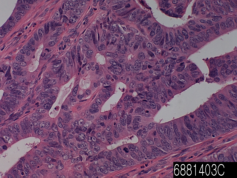
Cat. No. 6881403C
No. of samples 1
Description rectum adenocarcinoma PD
Age/Sex : 55/M
Price 197 EUR
260 USD
170 GBP

Rectum adenocarcinoma PD

Product Related Literature

Seen in young women in general, and have a better, VGA high differentiated adenocarcinoma villoglandular villoglandular papillary adenocarcinoma, and papillary villoglandular adenocarcinoma, has been shortened, as well as other cervical adenocarcinoma Villoglandular, in relation to cervical cancer, prognosis is a rare type of cervical cancer. Exhibition compression replaced papillary adenocarcinoma, tumor, normal lung tissue and form papillary structure complex, is the organizational form of lung cancer that is malignant cell growth diagnosis of destructive. The name of the lesions, describes the microscopic appearance of it. It is covered with glandular pseudostratified columnar epithelium, it has a structure like nipple and (papilla) long fibrovascular core and its relation to the width, (such as villi). The earwax adenocarcinoma is a malignant tumor of the auditory canal gland of the ear canal. In the number of names used in the past, these tumors are rare. Earwax adenocarcinoma cylindroma, ceruminoma, otherwise specified, (NOS), cystic carcinoma ceruminous adenoid (ACC), and mucoepidermoid carcinoma ceruminous is included synonym.

All of the tumor soft tissue, ear canal benign gland, and is a bone involvement or /. It may be deployed in a flat surface epithelium covering the tumor, but usually, it does not come from the surface epithelium. Tumors are located in cells, solid, cystic, cribriform glands, the single-cell pattern. Be displayed tumor necrosis is rare it, but if it is present, it is a diagnosis of cancer. The same is true of perineural invasion. A core containing a prominent nucleolus, in order to identify, easy, nuclear pleomorphism is usually. There is usually increased mitotic figures and atypical form. There are usually areas of interstitial fibrosis. It can be seen in benign tumors, but ceroid is not seen (ear wax and ear wax), in malignant tumors. The specific function of each tumor type, you will be able to support the separation into mucoepidermoid type and adenoid cystic carcinoma. This, it directly, to exclude the tumor by extending the ear canal from the parotid salivary gland is especially important when dealing with skin cancer or visco-adenoid cystic. This may be removed by imaging studies or clinical. Otherwise, histologic differential diagnosis includes nerve endocrine tumor or middle ear (benign ceruminous gland tumor) earwax adenoma (middle ear adenoma).

Management of magnesium and intravenous calcium is due to research results were presented at the 2013 annual meeting of the American Society of Clinical Oncology What is a benefit with oxaliplatin in order to reduce the side effects of neuropathy in colon cancer.

In this study, FOLFOX – The magnesium sulfate patient of 353 people with colon cancer receiving adjuvant therapy (5-fluorouracil, oxaliplatin, and leucovorin) and was added 1 gram of calcium gluconate before and after oxaliplatin randomly I was assigned to receive either placebo or one gram (CaMg). In the test third group, patients received a placebo before and CaMg after oxaliplatin. It will be evaluated by both the physician and patient questionnaire, there was no difference between the groups with respect to cumulative sensory neurotoxicity either or acute neurotoxicity. Adenocarcinoma, may occur in higher mammals, including some people. These cancers appear as glands, you may have the secretion characteristics. You may not have the appearance of a clear gland in some cases they. Each gland, may not be used to secrete the same material, but the name of any secretion characteristics, called adenocarcinoma malignant appearance and gland form is attached. Glandular tissue and epithelium because it occurs widely in the body, in adenocarcinoma, it is possible to affect several organs. Primary cancers of the colon are adenocarcinomas, it is quite common adenocarcinoma in the lung.

The most common type of cancer of the pancreas is a adenocarcinoma of the pancreas. 85% of malignant tumors of all of the pancreas is a gland cancer. Pancreatic adenocarcinoma is a leading cause Fourth of cancer deaths in men and women in the United States. Estimates of the American Cancer Society 29,000 U.S. each year, has been diagnosed with about 28,000 die of pancreatic cancer and adenocarcinoma of the pancreas.

It is believed that about 20 to 40% of pancreatic cancer patients have a cancer that is completely included in the pancreas at the time of diagnosis. Surgical removal of the tumor, is recommended in this group of patients, this provides the best option for long-term survival. For an operation highly specialized, surgery of pancreatic cancer should be evaluated by a surgeon experienced in the treatment of this disease is very patient.

About 60% to 80% of patients, because it is invade surrounding tissue (spread outside the pancreas) cancer or pancreatic outside it has metastasized, locally advanced cancer has been discovered.

Rectum adenocarcinoma MD

Product name Rectum adenocarcinoma MD
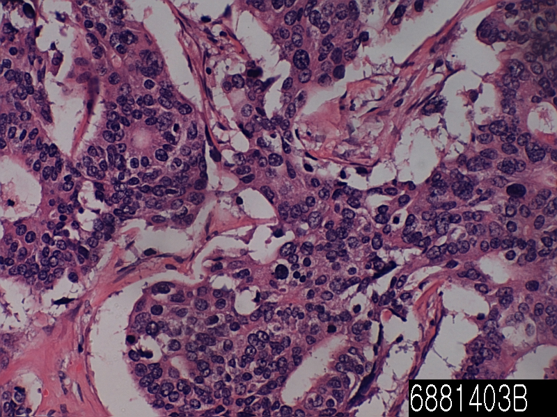
Cat. No. 6881403B
No. of samples 1
Description rectum adenocarcinoma MD
Age/Sex : 63/F
Price 197 EUR
260 USD
170 GBP

Rectum adenocarcinoma MD

Product Related Literature

The rectoprostatic fascia is a membranous partition in the lowest part of rectovesical bag. This split of the prostate and bladder of the rectum. It consists of a fibromuscular structure with several layers, which are fused together and cover the seminal vesicles. It is also called fascia Denonvilliers “, after French anatomist and surgeon Charles-Pierre Denonvilliers. Structure corresponds had rectovaginal fascia in women. At postoperative transsexual women, the vaginal cavity created by him. Rectoprostatic in the rear bumper also inhibits proliferation of prostate adenocarcinoma due to the invasion of the rectum is more often than is the invasion of other adjacent structures.

The objectives of the follow-up are to be diagnosed at an early stage or metastasis or tumors that develop later, but did not originate from the original cancer. The U.S. National Comprehensive Cancer Network and American Society of Clinical Oncology provide guidelines for the monitoring of colorectal cancer. A medical history and physical examination is recommended every 3 to 6 months for 2 years, and then every 6 months for 5 years. Carcinoembryonic antigen blood measurements follow the same time, but it is recommended only in patients with T2 or more lesions, which are candidates for intervention. A CT-scan of the chest, abdomen and pelvis can be considered for the first year, 3 years for patients who are at high risk of relapse (e.g., patients who are poorly differentiated tumors or venous or lymphatic invasion) and are candidates for therapeutic operation (to be treated). Colonoscopy can be done after one year unless it can be done during the initial braking by preventing mass, in which case it should be performed after 3 to 6 months. If villous polyp, a polyp> 1 centimeter or high grade dysplasia is found, it can be repeated after 3 years, then every 5 years. For other offenses, a colonoscopy can be repeated after 1 year. Routine PET scan or ultrasound, chest X-ray, complete blood count and liver function tests is not recommended. These guidelines are based on recent meta-analyzes showing intensive surveillance and close follow-up can reduce the 5-year mortality rate from 37% to 30%.

Cytopathology is a branch of pathology that studies and diagnoses diseases on the cellular level. Discipline was founded by Rudolf Virchow in 1858. A common application of Cytopathology a smear is used as a screening tool for the detection of pre-cancerous cervical lesions, and the prevention of cervical cancer. Cytopathology is also commonly used in the investigation of thyroid disorders disorders including sterile body cavities (peritoneal, pleural and cerebrospinal), and a wide range of other body sites. It is commonly used to aid in the diagnosis of cancer, but it also helps in the diagnosis of certain infectious diseases, and other inflammatory conditions. Cytopathology typically used for samples of the free cells or tissue fragments, in contrast to histopathology and studies whole tissues. Cytopathologic tests are sometimes called smear tests because samples can be smeared on a glass microscope slide for subsequent staining and microscopic examination. However, sputum samples can be prepared in other ways, including cytocentrifugation. Miscellaneous Pap tests may be used for diagnosis of cancer. In this sense, it is called cytology smear. Cytopathology is often less accurately called cytology, which means “the study of cells.”

Various normal function of cell growth, metabolism, and division may fail or operate in unusual ways and cause disease. Cytopathology is best used as one of the three instruments, the second and third are physical examination and medical imaging. Cytology can not be used to diagnose the condition of the patient and the replacement operation, to obtain a larger sample. An example is the thyroid FNA; many benign conditions can be diagnosed with superficial biopsy and the patient can return to normal activities immediately. If the malignant disease is diagnosed, the patient may be able to launch the radiation / chemotherapy, or it can have an operation to remove and / or stage of the cancer. Some tumors may be difficult to biopsy as sarcomas. Other rare tumors my be dangerous to biopsy as pheochromocytoma. In general, fine-needle aspiration can be done anywhere, it’s safe to put the needle, including liver, lung, kidney, and superficial masses. Many doctors are not trained to perform fine-needle aspiration biopsies correctly and then when they do not get diagnostic material believe that cytology is not useful. Proper technique takes time to master. Cytotechnologists and cytopathologists can help doctors by going to the procedures and assist with collection techniques. A “speed reading” is a look under the microscope and tell your doctor if sufficient diagnostic material is obtained. Sputum samples must be well prepared, so that the cells are not damaged. Sometimes more information about the model is helpful. Immunohistochemical staining and molecular testing may be performed, particularly if the sample was obtained using a liquid-based cytology. Often the “Reflex” test is performed, such as HPV testing of an abnormal Pap test, or flow cytometry of lymphoma model.

Rectum adenocarcinoma WD

Product name Rectum adenocarcinoma WD
Cat. No. 6881403A
No. of samples 1
Description rectum adenocarcinoma WD
Age/Sex : 44/M
Price 197 EUR
260 USD
170 GBP

Rectum adenocarcinoma WD

Product Related Literature

Cancer is the medical term for the most common types of cancer occurring in humans. To put it simply, the cancer is a cancer start in tissue that arise from cells of ectodermal and mesodermal origin embryonic mesoderm to the general, the surface of the inside or outside of the body line. In particular, were obtained from the epithelial cells to be estimated changed or damaged to the extent that such cells are transformed in their genome cancer tumor tissues, and began to show the characteristics of the abnormal malignant.

Long-term cancer refers to placing a small and localized prostate cancer that do not penetrate the epithelial membrane at the boundary of the cancer cells from normal cells adjacent still. Rather than invasive cancer in advance, CIS is a pre-malignant human. Until it begins to penetrate and infiltrate through the structure of adjacent / or other basement membrane with them, in the case of almost all of the CIS, we will continue to grow and develop. After the invasion happened, they do not think the CIS lesions longer, but it is invasive cancer really. Before and metastasis frank invasion will develop, it is not possible for you to be removed in the target treatment other areas of some damage surgical resection completely, cryotherapy, or laser ablation,, cure rate CIS 100% I approaches. In some cases, as it may histological characteristics and cytological appear more normal again assumed system lesions gradually, it is a low-grade tumor. Biologically, this can lead a less often aggressive, to slow tumor growth. In fact, the emergence of local tissue architecture and components of the Local Site CIS may be normalized to a point that does not meet the required consensus requirements to be classified as a cancer longer transformed cells eventually. Therefore, this anomaly is not allowed as a true cancer. The change of these, decrease the normal area, and I’m with the volume of place or abnormal /. The number that can not be ignored or some cases, the abnormal cells / tissue, may disappear the region obtained including the organization that looks normal only in full.

In many cases, the process is referred to by the pathologist as regression and specialist of the CIS lesion. Without connective tissue, associated, which is formed as a result of fibrosis apoptosis and necrosis often effective, involution CIS is gradual transformation of malignant tumor benign localized region of tissue near-normal or normal I bring. Regression occurs after prolonged exposure to changes in the strength and quality of immunological stimulation and environment most often and / or. In the example a very common, it is a regression of the disease of the CIS some located in the segmental bronchi and the main center of the lung. Invasive lesions of many conventional in the case of squamous cell carcinoma, reduced regression to the following long-term exposure of cells and tissues affected by the stimulation of the initial environmental carcinogen with what you see in abstinence after a long period of time from smoking like this often. A common example of a relatively different, causing regression of cervical CIS lesions, it is end is driven immunologically of human papillomavirus HPV by epithelial cells that have been converted of the cervix.

The (meaning straight intestine from Latin rectum) rectum, in mammals, are linear and the last part of the colon in the gut of the other. Human rectum is about 12 cm, starting at the level of the sacral promontory, and sacral third according to the definition (the end of the S-shaped colon) colorectal S-shaped junction is used. Its diameter is the same as the S-shaped colon at the start, it has expanded to its end, to form the rectal ampoule. Depending on the definition that is used again, it ends at the level of the dentate line (or at the level of the puborectalis sling) anal ring. In humans, the anal canal is followed in front of the digestive tract rectum ends with anal verge.

There are a number of rare subtypes undifferentiated, of undifferentiated carcinoma. (Including elongated cells similar to cancer of the connective tissue), a (huge, strange giant cell carcinoma spindle cell carcinoma: a lesion pseudo sarcoma component is included is included in some of the well-known including the polynuclear cells), and cell carcinoma huge (mixed spindle sarcomatoid carcinoma). Includes a cellular component and a huge spindle cell and / or polymorphism cancer is at least 10% of the component characteristic of a wide variety of cells (eg, squamous cell carcinoma and adenocarcinoma or /) of the species. In rare cases, tumors are facing cancer and sarcoma truth including sarcoma of the pulmonary blastoma that may not contain a component, similar.

In a logical way in order to obtain information about the degree of invasion and metastasis size and physical / clinical examination, cells and tissues, surgical technique, tumor staging of cancer, pathological of imaging studies and clinical examination I refers to the process of combining the inspection. Cancer, usually placed Roman numeral. Tumors smaller stage II cancer and I the classification stage, most were confirmed when and that is spread only in local structure and / or has been found. While it has spread in the body structure usually, lymph nodes, and / or tissue have been found, III stage cancer metastatic disease in the blood to the organs of tumor stage IV, distant sites, or organization.

Colon adenocarcinoma metastasis to liver

Product name Colon adenocarcinoma metastasis
Cat. No. 0062000A
No. of samples 1
Description colon adenocarcinoma metastasis
Age/Sex : 41/F
Price 197 EUR
260 USD
170 GBP

colon adenocarcinoma metastasis to liver

Product Related Literature

In metastatic disease or metastasis, is the spread of cancer to some non-adjacent organ or another organ or a portion of one. Was thought to be capable of infection or malignant tumor cells to metastasize formerly (abbreviated Mets) transition is generated in this way, a new instance of the disease is called, but since it is, further research is revised will be. Home transition is a Greek word meaning στάσις, stagnation, “accommodation” μετά, and, “Next”, “deviation” from the meta. Plural form is a transition.

Cancer can occur in the cells of the damaged cells genetically gradually be generated by proliferation unorganized controlled. The non-proliferation this control, cell division generates the primary tumor. Cell is a tumor undergoes metaplasia anaplaziya is followed by the results of the malignant phenotype, finally dysplasia. Extravasation of the position of the second for tumor formation is followed, this allows the blood vessels of malignant phenotype in blood circulation.

After that they move through the (circulating tumor cells) blood flow to the tissue and other parts of the body is possible, some cancer cells may acquire the ability to penetrate the vessel wall and lymph / or. This process is known (respectively), such as lymph or hematogeneous rating as well. When the tumor cells come to rest in other places, they continue to re-enter multiplying the walls and blood vessels, thereby forming a detection tumors clinically different in the end. This new tumor is also known as a tumor (or secondary) metastatic. Transition is one of three features of malignancy (contrast benign tumors). Varying degrees (e.g., basal cell carcinoma is not transferred almost) is most tumors, and metastasis.

When cancer cells metastasize, the new tumor is called metastasis or secondary cancer, the cells are the same as the primary tumor. Breast cancer, with metastases to the lungs of the secondary tumors, for an example, instead of the abnormal cells in the breast, the unit is made from the lungs for abnormal cells. Thereafter, tumors of the lung is called metastatic breast cancer, not the lung. Cancer cells to leave the tumor site of the original shifted to other parts of the body via the blood, where metastasis, by direct or expansion, is a series of complex steps of the lymphatic system. To do so, malignant cells is connected to the break away from the primary tumor, separating the tumor from the surrounding tissue, and degradation of extracellular matrix (ECM), about the make-up protein. The proteolysis of these cancer cells can be destroyed and escape ECM. In the trend at a rate higher than expected from statistical randomness, spread to tissue or organ specific, the position of the transition is not a coincidence and different types of cancer always. Breast cancer, for example, tend to bone metastasis, the lungs. This feature, chemokines are thought to be mediated by soluble molecules signals, and converts the growth factor β such. By the action of a class of proteins known as metastasis filter dozen is known, the main body to resist the transfer by a variety of mechanisms.

Amoeboid movement and population mobility mesenchymal-type exercise: human cells have three types of exercise. Cancer cells switches the different movements often opportunistic. Either stop, by blocking the required several steps of the movement of both types of either or in some way at least, researchers some cancers, treatment can slow the spread of cancer I hope that you find. Review the conditions required for cancer metastasis, called tumor angiogenesis, researchers cancer found that one of the most important events are necessary for the growth of new vascular network. This, it is possible inhibitors of angiogenesis may prevent the occurrence of metastases were found.

There are different types of cells several essential for tumor growth. Specifically, endothelial progenitor cells, is very important for the growth of the cell population of tumor vessels. Along with the fact that endothelial progenitor cells that is important for angiogenesis and metastasis, this finding was published in the gene development and science magazine. The importance of endothelial progenitor cells in metastatic tumor growth, angiogenesis and was confirmed by the recent publication of cancer research. In this paper ingenious, that it can be labeled with inhibitor 1 DNA binding endothelial progenitor cells. This new discovery is that it also was able to monitor the endothelial progenitor cells from bone marrow into the blood of tumor stroma, researchers, it was incorporated into the tumor vasculature. This finding of endothelial progenitor cells involved in tumor vasculature, vascular development in metastatic tumors and security demonstrates the importance of this cell type. In addition, removal of endothelial progenitor cells in the bone marrow produces a significant decrease in vascular development and tumor growth. Thus, endothelial progenitor cells, is very important in a new therapeutic target of current and tumor biology. In the process of cell movement in the base of the molding transfer, NFAT transcription factors are involved in breast cancer in particular. Indeed, NFAT5 and NFAT2 are migratory birds in breast cancer and Puropuro invasive, it is an inhibitor of NFAT3 cell movement. Adjusting lipocalin 2 to increase the invasion of breast cancer cells, and regulates the expression of the ligand and TWEAKR, NFAT1 inhibits the NFAT3 lipocalin 2 expression slowed cell infiltration.

The liver, an important organ in animals and some other vertebrates are present. It has a wide range of functions, including detoxification, protein synthesis, and production of bio-chemical and necessary for digestion. It can be used in the short term new technologies liver dialysis, a method of compensating required for survival, the lack of long-term liver function, there is currently no liver. It plays an important role in metabolism, glycogen storage, disintegration of red blood cells, plasma protein synthesis, hormone production, and the body has a number of functions in the body, including detoxification. It is located below the diaphragm in the abdominal-pelvic region of the abdomen. It produces alkaline compound to help digestion by emulsifying bile, the lipid. Many of them, small is necessary for the normal function of life, organization highly specialized liver is to regulate a wide range of mass biochemical reactions such as decomposition and synthesis of complex molecules.

Colon adenocarcinoma mucinous

Product name Colon adenocarcinoma mucinous
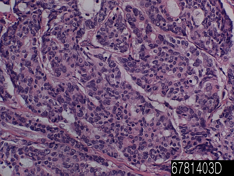
Cat. No. 6781403D
No. of samples 1
Description colon adenocarcinoma mucinous
Age/Sex : 44/M
Price 197 EUR
260 USD
170 GBP

Colon adenocarcinoma mucinous

Product Related Literature

Mucinous cyst adenoma is a type of unification of tumor cyst adenoma. To those associated with invasive cancer, those not associated with invasive cancer – pathologist classifies intraductal papillary mucinous tumors (IPMNs) into two main groups. This separation is an important prognostic significance. Patients with intraductal papillary mucinous tumors are surgically removed that are associated with invasive cancer while have a poor prognosis, patients with surgical resection intraductal papillary mucinous tumor without invasive cancer related, I have an excellent prognosis. Intraductal papillary mucinous tumor without invasive cancer and the corresponding can be sub-categories into three groups further. They are IPMN and IPMN with high-grade dysplasia, and IPMN, dysplasia of low-grade, and moderate dysplasia. Since it is believed to be shifted from moderate dysplasia IPMNs, high-grade dysplasia or low-grade dysplasia, this classification, it is more important than isolation of cancer IPMNs connected by IPMNs each without invasive cancer it is but, IPMN is useful classification is associated with invasive cancer.

Intraductal papillary mucinous tumors, can come to the attention of the clinical in a variety of ways. Abdominal pain, nausea, the most common symptom is vomiting. (Skin and eyes caused by obstruction of the bile duct become yellow) jaundice patients with the most common symptoms to come to medical attention, weight loss, and there is acute pancreatitis them. That it is to establish the diagnosis more difficult, the signs and symptoms of these are not specific for internal papillary mucinous tumor. Therefore, in many cases, the doctor tells the additional checks. If you have any reason to believe that it may patient has internal papillary mucinous tumors, he or she, the physician can confirm that suspicion by using one of the imaging techniques. These include (MRCP) (EUS), magnetic resonance cholangiopancreatography computed tomography (CT), endoscopic ultrasound. These tests, to clarify the expansion of one of the branches of the pancreatic duct or pancreatic duct. In some cases, fine needle aspiration (FNA) biopsy can be obtained to confirm the diagnosis. Fine needle aspiration biopsy can be performed through the skin using a needle guided by computed tomography or ultrasound that is performed through the endoscope in the endoscopic ultrasound examination, or can.

(Small space or cavity) IPMN cysts of the pancreas. These cysts is seen in CT (X-ray computed tomography). However, many pancreatic cysts are benign (see the disease of the pancreas). Before developing the (asymptomatic patients) symptoms, increase in number of patients are diagnosed They are now. Another reason, when it is scanned in the (X-ray), in these cases, (incidentally) patients discovered accidentally damaged pancreas. I will be done to treat the IPMNs occasionally to 6% of patients who underwent pancreatic resection.

Scientists Sol Goldman Pancreatic Cancer Research Center reported in the 20 issue July Science Translational Medicine at the Johns Hopkins University, they are genes that can be used to distinguish harmless from gun cysts of the pancreas I developed a test based. Test might help you avoid surgery patient harmless cyst is unnecessary. And his colleagues Bart Vogel, was found to be (intraductal papillary mucinous tumor) has a mutation in the gene of GNAS GNAS and / or pre-cancerous cyst almost all of the pancreas. Researchers have tested a total of 132 intraductal papillary mucinous tumors for mutations in GNAS and KRAS. Almost all was or both mutation, of GNAS of KRAS. Harmless cysts and serous cyst adenoma as had KRAS mutations or GNAS, researchers then feel safe. Before it is possible to provide wide genetic testing, many patients should be tested further.

Thus, treatments of choice for the main channel IPMNs resection is the probability of approximately 50% of malignant tumors. Side branch IPMNs can be seen on a regular basis by the MRI or CT, but sometimes, cut at a rate of 30% of malignant tumors in the resection of these tumors eventually. About 80% in malignant, there was no 85% of the malignant disease survival after resection of IPMN five years, lymph nodes, and there is no circulation at all and 0% growth of malignant lymph nodes. It may include to move the body and tail of the pancreas removed whole or pancreas (pancreatic tail) of the (total pancreatectomy) below, to remove the head of the pancreas (pancreaticoduodenectomy). In some cases, surgery may be performed using minimally invasive techniques such as robotic surgery or laparoscopy.

Colon adenocarcinoma PD

Product name Colon adenocarcinoma PD
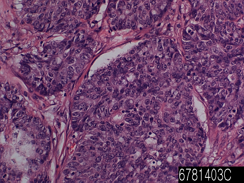
Cat. No. 6781403C
No. of samples 1
Description colon adenocarcinoma PD
Age/Sex : 37/F
Price 197 EUR
260 USD
170 GBP

Colon adenocarcinoma PD

Product Related Literature

Gland cancer is a carcinoma derived from glandular tissue. The epithelial tissue, skin, glands, organs and cavities and body line that is not limited to the surface layer of a variety of other organizations. Epithelium can be derived from embryology from mesoderm ectoderm and endoderm. So that they have a secretory properties, necessarily, it is classified as adenocarcinoma cells need not be part of the gland. Poorly differentiated adenocarcinoma can be differentiated adenocarcinoma, but they tend to resemble the glandular tissue that they are derived from. By staining the cells biopsies, pathologist determines whether the other type of cancer tumor or adenocarcinoma. Adenocarcinoma, the spread of glands in the body, in nature, and as a result, is likely to occur in many tissues of the body. Each gland is not able to secrete the same substance, but as long, as is the exocrine function in a cell, is considered the shape glands and malignant called adenocarcinoma it. Endocrine glands, such as VIP-OM, such as pheochromocytoma and insulinoma, tumor rather than not referred to as adenocarcinoma in general, it is called a neuroendocrine tumor frequently. No abnormal glandular tissue, in the case of benign, the adenoma says. Normal, benign adenoma is not rare transition If you do not invade other tissues. Adenocarcinoma malignant, metastatic given enough time to invade other tissues, and so often.

Symptoms of this type of cancer can be pain in the abdomen, weight loss, fatigue and weakness. Tumor size is increased, you may want to block the passage of food with crushed. There is a possibility that the tumor can lead to blockage. There is an obstacle when the intestine is blocked, you can not move anything to itself. This causes the pain to severe nausea and vomiting.  Can cause perforation tumor. In other words, the entire contents of the small intestine spill into the abdominal cavity when. Sudden severe pain, nausea, symptoms of perforation is vomiting. However, it is rare in itself.  Tumor starts bleeding in the gut sometimes. Slow bleeding so that the number of red blood cells is low. In weakness and fatigue, and as a result, this is also known as anemia. there is a possibility that rapid bleeding stool becomes black, digestion, causing slowed from the blood. This will allow you to feel dizzy or patient even if unconscious. If the process is a cancer, since it represents is described later, the diagnosis of cancer is known as adenocarcinoma NOS cancer, or otherwise stated is important that. But is determined by the organization they words are getting to tumor cells, ie, tissue origin is very useful for the projection of the process liquid, the adenocarcinoma of the colon, it has the treatment of ovarian cancer and prognosis, various You. It is shown in the diagnosis and particularly frequently, adenocarcinoma can be demonstrated by a pathologist using immunohistochemistry frequently. Since it is possible to find the primary site cancer is also called cancer of unknown primary.

The majority of colon cancer is adenocarcinoma. This is because has a plurality of inner gland tissue of the large intestine. Tend to be simple and tubular in appearance with a mixture of water absorption cells and goblet cells that secrete mucus normal colon glands. For secreting substances into the lumen of the large intestine, these glands are called glands, they are fluid the material. The purpose of these glands is double. The first is to absorb water from the feces back to the blood. In order to lubricate the feces dehydration now, the mucus in the large intestine lumen second aim. This is very important because that may lead to degradation of the stool column the establishment of lubrication of feces through which it passes into the rectum.

The glands of these, if you have received a series of changes at the genetic level, they will move in a predictable way these to be moved from benign colon cancer invasive, malignant. The study their paper, and Vogel, many more “Lessons from colon cancer is genetic,”. This suggests that colon cells lose the APC tumor suppressor gene, and a small polyps. Then, small polyps and that of K-Ras is activated, a benign adenoma. Not be attached to the end of it in the adenoma “cancer” does not suggest that it is of the benign and malignant adenocarcinoma. Gastroenterologist is using colonoscopy to find and remove polyps and adenomas of these you will not be able to continue to acquire genetic changes that they lead to invasive adenocarcinoma. And Volgelstein. Loss of p53 and DCC genes suggests that brought adenocarcinoma malignant further.

Grossly, the table seen in a different color than the surrounding tissue will show people. Often, bleeding from a tumor is evidenced by a tendency to increase the tumor vessels therein in a random manner through the secretion of a number of pro-angiogenic factors such as VEGF. The Histologically, the tumor was similar, are classified original structure as they were differentiated. Tumor cells that have lost the similarity of any original tissue structure shaped appearance and both are shown as poorly differentiated tumor cells. Tend to have a large nucleus with a prominent nucleolus grade, malignant tumor regardless. In addition, there is a significant increase in the incidence of cell division or mitosis.