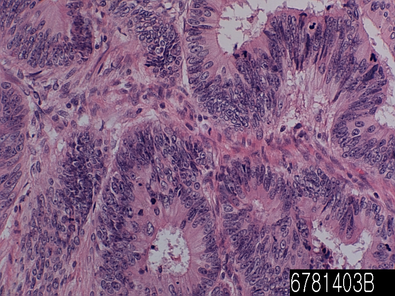
Colon adenocarcinoma MD

Product name Colon adenocarcinoma MD
Cat. No. 0062000A
No. of samples 1
Description colon adenocarcinoma MD
Age/Sex : 56/M
Price 197 EUR
260 USD
170 GBP

Colon adenocarcinoma MD

Product Related Literature

As a consequence of mutations in that Wnt signaling pathway to increase the signaling activity artificially most commonly, colon cancer is a disease derived from epithelial cells in the mucous membrane of the colon or rectum of the gastrointestinal tract. Mutations can be acquired or inherited, to generate stem cells in intestinal crypts most likely. Mutant gene is the APC to produce APC protein most often in colon cancer gene of all. In the “brake” of accumulation catenin protein, a non-APC, tail – – tail APC protein catenin accumulates to high levels in the nucleus move to (drive), a general importance to update the system and bind DNA Sometimes, expressing cell differentiation, can lead to cancer in inappropriately high level when you activate the transcription of a gene. APC is mutated in the majority of certain types of cancer, colon cancer, cancer, blocking mutations in other genes with similar functions or decomposition, its tail – tail for catenin is a (CTNNB1) – NKD1 or, AXIN2, TCF7L2, ADR as AXIN1 catenin mutation is increasing.

Beyond the defects in Wnt signaling-APC-β-catenin signaling pathway, must be performed to convert the cancer cells other mutations. The monitor cell division usually, if there is defect in Wnt pathway thereof, P53 protein production differences, kill cells by TP53 gene. Finally, the cell line, we get the organization turns TP53 mutations and gene from adenoma to invasive cancer. Delete (in colorectal cancer (sometimes mutations are not a protective another protein named BAX is p53 gene coding), DCC and TGF-tail is a apoptotic protein disabling other colorectal cancer in general ). And has a mutation that inactivated at least half of the TGF-tail colon cancer. In many cases, a protein called Smad DCC followed when it is not TGF-tail excluded people, is the deletion of chromosomal segments in colorectal cancer.

Several genes is a cancer gene – They are over-expressed in colon cancer. For example, a protein gene encodes, usually, PI3K KRAS, and RAF may acquire a mutation that results in the activation of cell proliferation and stimulates cells to divide in response to growth factors. General primary KRAS mutations lead to borderline lesion or self-limiting hyperplasia, chronological order of mutations is important sometimes, if it occurs APC mutation after the previous, in many cases, to progress to cancer. PTEN, tumor suppression, can usually inhibits the PI3K, disabling and sometimes mutations.

I made it clear that colon cancer is ultra-mutation, comprehensive analysis, genome-wide can be separated clearly in tumor types not super mutation. In addition to the above inactivating mutation for the gene that contains a super-mutation and ARID1A, SOX9, ATM CTNNB1, FAM123B mutations also sample and oncogenes. Genetic event that displays a mutant of BRAF and, TGFBR2, MSH3, MSH6, SLC9A9, TCF7L2 ACVR2A, progression through a separate set of super-mutant tumor. The common theme in both types of tumors among these genes, it is the participation of TGF-tail signaling pathway, and WNT lead to increased activity of the major factor MYC, of ​​colon cancer in order. Usually, it has been reported from the analysis of the tissue taken by biopsy or surgery in the histology of the tumor. Usually, a description of the grade and type of cell is included in the pathology report. The most common type of cancer of the colon cells are adenocarcinoma accounts for 95% of the cases. Common type of other less, includes lymphoma and squamous cell carcinoma. Tend to be meaning ekzofitichen, that the tumor grows outward from the part of the intestinal wall tumors (cecum and ascending colon) right. Not causing the blockage of feces very rarely, this is symptoms of anemia, such as such. Tumor on the left side, allowing you to make is very similar to the ring napkin, which allows that a butterfly has to offer for flights caliber thin tend to be circumference and, Yes.

It is a malignant epithelial tumor of glandular epithelium from adenocarcinoma, colorectal mucosa. Wall, it penetrates into the penetration of the muscle layer of the mucosa muscularis propria and submucosa from there. Tumor cells were described to hold the lumen pluristratification, multiple, irregular tubular structure, the stroma was reduced (the side of the “parallel”). Mucus (colloid) adenocarcinoma, poorly differentiated – was entering the stroma to generate a large pool of (space of “empty” optically) mucus / colloid, sometimes, tumor cells are mucus and discohesive. When remaining in the tumor cells, the fluid pushes the core to the outer periphery – “a signet-ring cell”. Depending on the predominant pattern of mucosecretion and polymorphism gland architecture, cell, be presented three times of differentiation can be adenocarcinoma: well, poorly differentiated and moderately.

Details of histogenesis of FA is still unknown, highly discussion. Usually in the heterogeneous than peritumoral, adenocarcinoma, and due to the malignant transformation of primitive cells that can exhibit the characteristics of goblet cells cell differentiation Clara, II lung alveolar cells, cell surface bronchioles, or bronchial gland cells, possible. High-grade top FA, poorly differentiated variants have been described, it was moderately differentiated tumors or enough. It is possible to find FA similar to tissue is mixed with a collection of primitive osteoblast-like cells. The presence of cell blastomatous exacerbate prognosis significantly, in this case, tumors of two phases can be classified as a form of pulmonary blastoma. The FA, may occur in combination with adenocarcinoma other forms of lung cancer, other embodiments in particular. In addition, there seems to be a clear link implementation of lung cancer. I reported the case of synthetic small cell lung cancer cells that are similar to those of carcinoid tumor components and the FA are involved, and show the complexity and diversity unique tissue formation of lung cancer and cell differentiation.
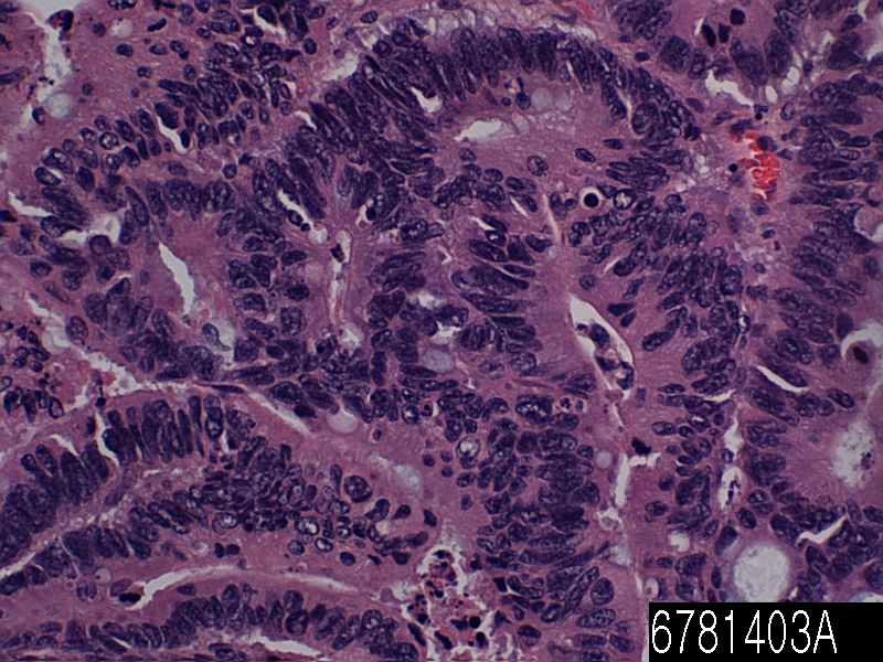
Colon adenocarcinoma WD

Product name Colon adenocarcinoma WD
Cat. No. 6781403A
No. of samples 1
Description colon, adenocarcinoma
Age/Sex : 48/F
Price 197 EUR
260 USD
170 GBP

Colon adenocarcinoma WD

Product Related Literature

In addition, in colon cancer known as cancer or colon cancer of the colon, cancer, are in addition to uncontrolled growth of cells in the colon or rectum or is (colon). The genetic analysis of colorectal tumor essentially, the same genetically, indicating that it is a cancer. Typically, symptoms of colorectal cancer, would include rectal bleeding that is associated with changes in bowel habits and weight loss, sometimes anemia. As a fraction of the cases hereditary disease underlying are involved, colon most cancers can be attributed to age and lifestyle. Usually begins in the intestinal mucosa, and if not treated, the lower, which in turn can be converted into the muscular layer through the intestinal wall. Screening and is effective in reducing the risk of death from colon cancer, it is recommended starting at age 50, people will continue until 75 years old. Usually, colon cancer, which is localized be diagnosed by colonoscopy or S-shaped colonoscopy. Cancer in the wall of the intestine, while the cancer has spread widely in the body, curing is not cured typically often surgically, control, attention to the durability of the human using chemotherapy then , I have improved the quality of life. Colorectal cancer is a cancer that is most frequently diagnosed in the three largest in the world, but the developed countries is common it. About 60% of cases are diagnosed in developed countries. It is estimated that that all over the world in 2008, new cases of 1.23 million of colon cancer will be diagnosed clinically, it killed 608 million people.

Symptoms and signs of colon cancer is dependent on the location of the tumor in the colon whether it has spread to other parts of the body. Traditional signs include: worsening of constipation, blood in feces, reduction of gauge stool, loss of appetite, weight loss, and anyone over the age of 50 nausea and vomiting. Rectal bleeding while, anemia is a feature of the high risk of Lowel practice on if bleeding is usually in those over the age of 50. People with (ulcerative colitis and Crohn’s disease) and inflammatory bowel disease, the risk of colon cancer is high. Long human risk is greater than had worsening severity of inflammation and disease. It is recommended for prevention and high-risk group of these in colonoscopy and regular aspirin. People with inflammatory bowel disease, accounting for less than 2% of colon cancer cases every year. 2% is a colorectal cancer 8% in ’10 after 20 years in those with Crohn’s disease, 18% after 30 years. About 16 percent of developing colon cancer or precursor cancer and after 30 years in those with ulcerative colitis.

Those having a family history of primary more than two, the risk of increased two to three times the disease, which will be explained about 20% of the group of all cases. Number of genetic syndrome is associated with rate of colon cancer in high. The most common of these is (Lynch syndrome or HNPCC) hereditary non-polyposis colon cancer, which is present in about 3% of those with colon cancer. Syndrome of other highly relevant include: familial adenomatous polyposis and Gardner’s syndrome is the responsibility of one percent of the cases that are occurring almost always cancer (FAP) is. Most of the deaths from colorectal cancer that is associated with metastatic disease. Transition associated with cancer Colon, one – the gene that appears to contribute to the likelihood of metastatic disease, I was isolated. This is a transcription factor that affects the expression of hepatocyte growth factor. This gene is linked to a distributed invasion of colon cancer in metastasis and tumor growth in mice and cell cultures, and distribution. It is also associated with response to treatment.

Change of () genetic variation of colon cancer general are also much more epigenetic changes. And others Vogel is as described, (together referred to as “driver mutations”) in, 1-5 tumor suppressor mutation with a mutation “passenger” and about 60 extra carcinogenic mutations in one or two only average colon cancer some. It has been well-studied tumor suppressor genes and oncogenes, will be described in the pathogenesis. However, in comparison, epigenetic changes in colon cancer is common, hundreds of genes are affected. For example, is about 22 nucleotides, small RNA species called micro-RNA,. It does not encode a protein (miRNA) or micro RNA of these, the gene encoding a protein “direct” They can be a decrease in their expression. The expression of miRNA These can be changed epigenetic. As an example, epigenetic changes consisting of methylated encoding meal -137 CpG island of the DNA sequence, epi that reduces its expression, which occurs 14% and 81% of colon cancer, and starts in Colorectal carcinogenesis often I appear in the colonic mucosa adjacent to the cancer usually is a genetic event. Change the tissue of interest that are associated with these cancers, called field defect. If you stop the meal -137, the purpose of this miRNA will be able to affect the expression of the gene of 500. Changes in the expression of the protein product of the gene, 2 to 20 times, even number of each change of MIR-137 expression levels leads to modification of mRNA expression of a target gene by decreasing frequently. Micro RNA other possibility is a number comparable target gene is high, more generally, to correct them arising from epigenetic defects, colon cancer region and also in the large intestine. MIR-342, 99% meal-124A, and 34B-MIR / C, and 93%, it has been blocked by methylation of the CpG island of the encoding DNA sequences in the primary tumor at a frequency of 86% each, these appear mucosa at a rate of 56% 59% respectively contain the normal adjacent, and 26%.

Small intestine malignant lymphoma

Product name Small intestine malignant lymphoma
Cat. No. 6495903A
No. of samples 1
Description small intestine, malignant lymphoma
Age/Sex : 20/F
Price 197 EUR
260 USD
170 GBP

Stomach signet ring cell carcinoma

Product Related Literature

Small intestine malignant lymphoma is part of the stomach under intestinal tract (small intestine) or the small intestine, and is where the colon is followed by most of the digestion and absorption of food is carried out. It receives pancreatic juice and bile through the control channel by heptopancreatic Oddi Spincter. In invertebrates such as worms, colon The term “gastrointestinal tract” is used to describe the entire intestine often. Information about the process is applied directly to the placenta of most mammals, but in this article, the human gut is about main. The primary function of the small intestine is the absorption of minerals present in the diet and nutrients. (The main exception to this rule is information of cattle for the digestion of a mammal similar to cattle,. Please refer to ruminants) that stuff digestion, pass the blood vessels in the intestinal wall in the process of diffusion I can. It is a site that is absorbed by the majority of nutrients from food ingested in the small intestine. The mucosa of the small intestine and inner wall, simple columnar epithelial tissue is lined. Structurally, it is covered with folds or wrinkles called annular folds are considered functional permanent wall mucosa and organs. Wrinkles that make it possible to stretch or considered non-permanent, they are different. Project of folds, circulares a part of microscopic finger of tissue called villi (Latin for “shaggy hair”). An extrusion finger known as microvilli epithelial cells of the individual. Features microvilli annular folds, and villi is to increase the amount of surface area available for absorption of nutrients.

Each villus has a network of fine capillaries and lymphatic vessels near the surface called lacteals. In the epithelial cells of the villi transport nutrients from the gut lumen to lacteals (lipids) and carbohydrates (and amino acids) of these capillaries. Absorber is transported via blood vessels other organs of the body that are used for the construction of a composite material such as a protein that is required in our body such. Food enters the large intestine without being absorbed and undigested. Shape and length are significantly different between species, but in the small intestine, quadruped, and found in all fish. In fish, for the length of one and a half times fish body, and relatively short, typically. Along serve to increase the entire surface of the body to digest pyloric vertically, often food, its length, it has a number of small bag like structure. Fish ileocecal valve not some boundaries between the small intestine and rectum such was observed only at the end of the intestinal epithelium.

Open the large intestine in quadrupedal of the ileocecal valve, so it will always be there. In comparison with four bony fish, the length of the small intestine is a long common, but it is a herbivorous animal, This is especially true in birds and mammals is higher than the proportion of reptiles and amphibians metabolism. The mucosa of the small intestine, in all vertebrates, in mammals only, increasing the surface, contains the fine folds have developed villi real them. Such distinctions are ignored one of the somewhat ambiguous boundary duodenum, jejunum, ileum, and even with people when discussing random in nature or anatomy, of other animals. Intestine as no bony fish, sharks, such as not, such as lungfish and sturgeon. Instead, some form of spiral casing of the digestive intestine Please connect the stomach rectum. This type of bowel cancer itself is relatively straight, but many times, there are dozens of turn by running along the inner surface of a spiral at times. This valve can improve both the effective length of the intestine and size substantially. Helix, tetrapod to intestinal mucosa of the small intestine as well as fish, in mammals. In the lamprey, probably because it requires little digestion, valve spiral very small their diet. helical valve instead of hagfish exactly occurred for substantially the entire length of the intestine is divided into regions digestion are different.

Lymphoma, T lymphocytes and B to form part of the immune system, protect the diseases and body infections, leukocytes, and blood cancer that occurs when it is alive or longer were separated more rapidly than normal cells It is expected there. It can be developed in other organs lymph nodes, spleen, bone marrow, blood, ultimately, lymphoma, form tumors. Typically, lymphomas present as solid tumors lymphoid cells. Treatment, in some cases may include chemotherapy, bone marrow transplantation and / or radiation therapy, and lymphoma histology, can be cured depending on the stage and type of disease. In many cases, it presents itself as an extension node (tumor), malignant cells of these are derived from a lymph node. As called extranodal lymphoma, as in this case, it can affect other organs. The site of extranodal, bone skin, brain, and intestines, is included. Be associated with lymphocytic leukemia derived from lymphocytes and lymphoma closely, but is contained (which is created by a process known as haematopoesis the blood cells) and bone marrow circulation generally static tumors generally do not form. There are many types of lymphoma there, in turn, lymphoma is a part of the group a wide range of diseases, called the blood tumor.

The current definition, “classification of lymphoma of human American and Hispanic Europe revised” and is the classification of the latest of lymphoma is a WHO classification published in 2001, was updated in 2008 was placed in a (REAL) in It is based on the foundation. By determining phenotype cell type (i.e., normal cell types are similar to most tumors), and a molecular cytogenetic characteristics, the system attempts to group lymphomas. There are three major groups: cells, natural killer cells and T-cell tumor. Group less common other I will be recognized. Are considered separately in the World Health Organization in but significantly abnormal, lymphocytes of B-cell lines mature, Hodgkin’s lymphoma, classification (and earlier), is recognized as a tumor now. After the diagnosis and treatment has been staged in front of the cancer. This applies where ever to the site and other local organizations, the cancer has spread to display. Staging is reported between class I and (limited) IV of (spread). Staging, and is performed as stage of the cancer, I will affect the treatment and prognosis. Ann Arbor staging system is used to the suspension of the NHL and HL in general. In this suspension system, I is a disorder localized in the lymph node, the presence of lymph nodes more than one II two, the spread of lymphomas on both sides of the diaphragm to the lymph nodes outside the organization, III can, and IV. PET scans or CT scan is used to organize the cancer.

Stomach signet ring cell carcinoma

Product name Stomach signet ring cell carcinoma
Cat. No. 6384903A
No. of samples 1
Description stomach, signet ring cell carcinoma
Age/Sex : 72/F
Price 197 EUR
260 USD
170 GBP

Stomach signet ring cell carcinoma

Product Related Literature

Ring squamous cell carcinoma is a histological variant of skin cancer (sometimes displayed as “squamous cell carcinoma signet ring”). As the name suggests, this is a subtype of squamous cell carcinoma, I is characterized by concentric circles made of vesicles that are substantially enlarged keratin, large vacuoles, the corresponding. These bubbles will rise to the extent that displacing the similarity of unique cell surface to the nucleus radical, “ring” of the cells to the plasma membrane when observed under a microscope. Signet ring cell carcinoma is a malignant epithelial tumor characterized by histological type of cell ring seal. It takes the form of adenocarcinoma, which is found in glandular cells of the stomach most commonly, but it also may occur in other areas of the body. For example. Interstitial testicular prostate, bladder, gall bladder, breast, colon, and ovary. In many cases, these cases and is inherited by (but not all) is caused by mutations in the gene CDH1 are some instances. Pattern of metastasis of gastric cancer cells with different printing than intestinal-type gastric cancer. When gastric cancer cells to transition the print, there is a tendency to create a Krukenberg tumor, spread to lymphocytic infiltration of ovarian peritoneal metastasis, lung and it.

The SCLC, may occur in a wide variety of combinations of histologic variants of other lung tissue containing complex malignant impurities very. If it is found in the form of a separate one or more of lung cancer such as adenocarcinoma and squamous cell carcinoma, malignant tumors, and then it was classified as (c in SCLC) mixed small cell lung cancer diagnosis and. Of SCLC, C non-small cell lung cancer, a subtype that is currently recognized. Synthesis SCLC is the current position, and are treated in the same manner as “clean” small-cell lung cancer, but recent studies have shown, the operation is able to improve performance in the early stages of this type of tumor .

Are as lung cancer and other symptoms. In addition, by the cell neuroendocrine origin of them, small cell carcinoma, and release substances that cause paraneoplastic syndromes such as Lambert-Eaton myasthenic syndrome such often. The main venue of small cell carcinoma, it is not a very rare outside of the pleural cavity and lung, it is called (EPSCC) small cell lung cancer. Outside of the respiratory tract, small cell carcinoma, can occur in the bladder cervix, prostate, liver, pancreas, or gastrointestinal tract,. Thought to consider a new case 1,000 per year in the United States that is histologically similar small cell lung cancer, the treatment of small cell lung cancer, it is used to treat EPSCC typically. Line initial therapy in general with etoposide and cisplatin. In Japan, the first-line treatment, transition to cisplatin and irinotecan.

Main spot of the skin when a Merkel cell carcinoma more. Small cell lung cancer is divided into two phases comprising (ES) stage a limited clinical pathological long and rich stage (LS). Tumors is shown only from the chest generally by step, whether or not it is that can be incorporated into the portal radiotherapy single total weight of the breast tumors practice is determined by the presence or absence of metastases. If it is limited to the lymph nodes near the lung and lung tumors one, it is said in general, and LS. If you have cancer spread beyond it, the ES is said.  In the case (in many cases, including paclitaxel cyclophosphamide, cisplatin, doxorubicin, etoposide, vincristine and / or) of LS-SCLC, combination therapy, I was administered together (RT) and concurrent thoracic radiotherapy.

Between 75% of people indicating the (CR) “complete remission” and 45% initial objective response rate very high between the observed using the LS-small cell lung cancer chemotherapy 90% from 60% ( It is defined as the disappearance of radiological evidence of clinical and all RR), of a tumor. Unfortunately, recurrence is the rule, the median survival is 24 months from 18 months. Under most circumstances, has spread widely in the natural history of the tumor at a very early stage, to respond dramatically in almost all cases, SCLC is or RT / CT, the role of little for surgery in 1970 this disease It is from the year. However, a recent study, when used before chemotherapy, in the case of (“very limited”) small asymptomatic, node-negative SCLC-san, surgical resection survival Improving was suggested. (“Adjuvant”).

The ES-SCLC, the combination chemotherapy, the standard treatment of radiation therapy, if temporary, shortness of breath, such as have faster than normal, and pain caused by bone metastases and liver, SCLC is for the treatment of brain metastases responses to whole brain radiation therapy were added only to alleviate symptoms such as.  Combination therapy is composed of cisplatin, cyclophosphamide, vincristine and carboplatin and, from a variety of agents. 30% of patients had a complete response to combination chemotherapy and 15%, response rate is also high for a wide range of disease among the majority of response objective of at least some.

However, in many cases, the response of ES-SCLC short time. If it occurs in the target having a small cell lung cancer complete response to chemotherapy, (PCI), is used to prevent the development of brain metastases often prophylactic cranial irradiation after. This treatment is very effective, but it can cause fatigue and hair loss. Adverse effect was not observed at all in the prospective randomized trial with 2 year follow-up neurocognitive almost. A meta-analysis of the PCI confirmation randomized trial, it provides a significant advantage for survival. Finally, small cells are very sensitive to radiation therapy and chemotherapy, platinum-containing agents, in particular, basic circuit. However, most people with median survival and disease relapse remains low.

Stomach mucinous adenocarcinoma

Product name Stomach mucinous adenocarcinoma
Cat. No. 6384803A
No. of samples 1
Description stomach, mucinous adenocarcinoma
Age/Sex : 55/M
Price 197 EUR
260 USD
170 GBP

Stomach mucinous adenocarcinoma

Product Related Literature

Cancer arising from epithelial cells, mucus cancer, (the main component of mucus) production of mucin and skin, and internal organs of the line fixed.  In the case where it is found in the skin, mucinous carcinoma is round often, but reddish, is located ulcer of the mass, the head and neck usually repeated for and sometimes grow,. (Mucinous tumors) or mucinous tumor is part of the surface epithelial stromal tumor group of ovarian tumors, accounting for approximately 36% of ovarian tumors of all. About 75% in benign, 10% in the border line, 15% are malignant. In rare cases, the tumor can not be seen on both sides, about 5% of primary mucinous tumors are bilateral. I have, a smooth mucosal epithelium similar to cervical epithelial cells and gastric epithelial cell cyst is usually small, benign tumors and mucus multilocular,. Boundary malignant mucinous tumors, have a solid region and nipple often. May also necrosis and bleeding.

Well, it is described that the sample as it needs further malignant tumors may be present solely rely mucus of ovarian tumors. Materials that give their names to those mucus-like, the main features of mucinous tumors are filled in is produced by goblet cells that secrete mucus that is very similar to the cells lining the normal intestinal this mucus It is a tumor that are. These tumors can become quite large, some are weighted km more than 25. Necrosis and loss of stratification and different type of cell, the normal structure of the organization: Cystadenocarcinomas (malignant tumor) includes a solid model of the growth of the characteristics of malignant tumor. Appearance, may seem like a colon cancer. A clear stromal invasion will be used for the differentiation of borderline tumors of cancer. Peritoneal pseudomyxoma, may be present as a result of ovarian mucinous tumors, but this is not the cause, of this state is the most rare state. A common cause of peritoneal pseudomyxoma is a mucin-producing tumor of the appendix. Usually, ovary mucinous tumors because it contains ovarian one, it means that it is possible to tumors is raised to another location, the presence of a portion of both ovaries and mucinous tumors are guaranteed further investigation You. Relative risk of 2.22 and 2.02 current smokers for smokers in the past: the risk of mucus tumor was associated with smoking significantly. Further risk is associated with the duration of smoking: RR 20 years was 1.44. Please refer to the article Tworoger SS of March 1 Cancer in 2008 using data from the Health Research nurse.

In vertebrates, it is mucous membrane secretions or that is derived from mucus is slippery. Normally, fluid mucosa produced by cells located in mucous glands. Cells in the mucous membrane to separate the rich product and water glycoprotein. It may come from a mixture of glands, including the mucosal cells and serous fluid also mucosa. Preservatives called mucins produced by submucosal glands and goblet cells mucosa, enzymes, immunoglobulins, inorganic salts, for example, this is a viscous colloid containing proteins and glycoproteins, such as lactoferrin. This fluid helps skin cells in the respiratory epithelium of mammals in gills amphibians, fish, digestive, urogenital, visual, auditory (pipelining), and to protect the system. For example, key features of one of the mucus is to protect against infectious agents such as viruses fungi and bacteria. Body of the average person produces about 1 liter mucus per day. Other invertebrates bony fish, hagfish, snails, slugs and produces the external mucus. It may be possible that in addition to play a protection against infectious agents, to provide protection against toxins produced by predators mucus such, to facilitate the movement and serves for communication.

The nose, particularly respiratory system including humans, a protective mucus aids, lung via the capture of particulate matter during breathing normally. While describing the secretions of the nasal cavity “nasal mucus” term “sputum” is the technical term for the mucus that is limited to the respiratory tract. Nasal discharge is generated by the mucosal tissue of the nose and mucous membranes that line (trachea, bronchi, bronchioles) airways produced by the specialization of submucosal gland epithelial cells and respiratory tract (goblet cells). Particles of small dust and infectious agents particulate pollutants, allergens, and bacteria, it is trapped in airway mucus or nasal adhesive and will enter the system is prevented. In constant motion of the respiratory mucus layer, this event will help to prevent the entry of foreign matter into the lungs during breathing in the oropharynx. explain why that happens to people who cough smoke filtered cigarette frequently. In order to increase the production of mucus, a natural reaction of the body. In addition, to prevent the epithelial tissue, such as drying and fluid aid in wetting of the intake air, and airway of the nose. even when it is dry, swallowed unconsciously almost, airway mucus and nose are always generated. To increase the production of mucus in the airways, it is a symptom of a common disease of many, such as a cold or flu. The mucus increased, may occur in inflammatory airways disease, respiratory allergies such, such as chronic bronchitis and asthma. The presence of mucus in the nose and throat is normal, but the amount of increase, you will need to be cleaned by blowing the nose or throat of phlegm discharge and preventing breathing comfort.

In general, the filtration function of air during inhalation, nasal mucus is clear and thin. Is at the time of infection, fluid, you can change its color as a result of the reaction of the body to viral infection and due to trap bacteria or green, and yellow. Green fluid coming from the heme group with an iron-containing enzyme myeloperoxidase secreted by white blood cells such as cytotoxic such defenses to respiratory burst.  In the case of bacterial infection, the bacteria cut the propagation of sinus and already congested in the medium nutrient-rich moist. Sinusitis is an uncomfortable condition may include an overload of fluid. The bacterial infection in sinusitis, usually, virus infection will respond to antibiotic therapy and discolored mucus that resolved without treatment. Sinus infection all, is a viral almost antibiotics is invalid and is not recommended for the treatment of a typical case.
The case of virus infection such as a cold or flu like this, a clear thin mucus, caused the rear of the nose and throat in the final stage of infection and the first stage,. So in order to correspond to (1 to 3 typically) virus, the body, as can mucus thick and yellow or green. Virus infection can not be treated with antibiotics, it is an important means for its misuse. Treatment usually is a symptom of the base, in many cases, it is sufficient to allow the immune system to fight the virus passage of time.

Stomach adenocarcinoma

Product name Stomach adenocarcinoma
Cat. No. 6381403A
No. of samples 1
Description stomach, adenocarcinoma
Age/Sex : 65/M
Price 197 EUR
260 USD
170 GBP

Stomach adenocarcinoma

Product Related Literature

Gastric cancer and gastric cancer, refers to cancer arising from any part of the stomach. Stomach cancer, cause death is bad is about 80 million people worldwide per year.Prognosis most patients, since exhibits a high degree of disease. Or is (produced without noticeable symptoms) or often asymptomatic, as well as symptoms is not a gastric cancer only (specific symptoms of non-specific only, stomach cancer, disease or irrelevant, other related in the first stage for) there is a possibility. By the time symptoms appear, cancer is the main cause (of the body, spreading remote, in part possibly other) has reached the stage to advanced also often (see below), and may transition relatively poor prognosis.

The most gastric cancer by Helicobacter pylori infection. This is why proven, but some foods such as pickles smoked food like this, and meat, and salted fish, nutritional factors that are associated with high risk. Nitrite and nitrate can be a compound which can cause stomach cancer in animals has been discovered, by certain bacteria, including H. pylori, is turned on in sausage. On the other hand, and, it is recommended to eat fresh fruits and vegetables that contain antioxidant vitamins, such as A and B to the American Cancer Society, they Mediterranean diet and, say, reduce the risk of cancer of the stomach, stomach and is associated with lower levels of cancer. Smoking increases the risk of developing gastric cancer significantly by in order to increase 82% for heavy smokers, risk 40% of current smokers is increased. In some studies, the esophagus has shown an increased risk of alcohol as well, gastric cancer caused by smoking occurs in the upper portion of the stomach mostly.

Other factors that are associated with increased risk is the genetic factors (hyperplasia, stomach hypersecretion), intestinal metaplasia, and autoimmune atrophic gastritis, pernicious anemia, Alzheimer Menetrier minutes. H. pylori is the main risk factor of 80% from 65 of cancer of the stomach, but only 2% of such infections. Pylori-induced mechanism of stomach by H. involves the virulence factors of action pylori or chronic inflammation of the H. CagA potentially. About 10% of patients showed a genetic component. Causal relationship has not been established, but several studies have correlated shows the spores of the consumption bracken, the incidence of cancer of the stomach. Gastric cancer, shows the male-dominated its incidence up to three men that are affected for all women. Estrogen, you may want to protect women from the development of this cancer form. A small percentage is believed to be a gene (forward references histopathology) very diffuse type gastric cancer. Hereditary diffuse gastric cancer, have been identified (HDGC) recently, research is in progress. However, treatment options and genetic testing is available for families at risk now. The International Cancer Genome Consortium effort to complete the genome map of gastric cancer.

Abnormal tissue found in gastroscopy is the biopsy gastroenterologist or surgeon. Thereafter, the fabric is sent to the pathologist for histological examination using a microscope to check for the presence of cancer cells. The biopsy and histological analysis of then, it is a certainty the only way to confirm the presence of cancer cells. It has been developed to increase the yield of open mucosal dye can condition gastroscope, various strengthen cell structure, to identify the region of dysplasia. Accompanied ultra-high magnification for visualizing cell structure to determine the region of dysplasia good Endocytoscopy. Further gastroscope conditions, such as optical coherence tomography such other tested for research purposes and the like.

State of some skin is associated with gastric cancer. It is known as acanthosis nigricans, in many cases, dark conditions, has been associated with cancer of the abdomen, such as gastric cancer, such as this for the thickening of the skin due to groin and axilla. The skin other symptoms of stomach cancer, Lesser · Trela ​​t symbol is also known as seborrheic keratosis and (darkening hyperplasia similar skin of the palm) tripe palm, will be a rapid development of skin lesions.  In order to check the anemia, complete blood count (CBC): Blood various tests may include the following, and is carried out. Also, stool test may be performed to test the blood in feces.

Cancer cells, is that when it is detected in a tissue sample, the next step is performed, or to grasp the extent of the disease. In various tests, it is determined cancer whether the spread, and if so, which parts of the body are affected. Gastric cancer is possible to order the tests or other tests, CT scans, PET, of the ultrasound endoscope in order to be able to spread to other organs liver, pancreas and near the stomach, doctor lung checks These areas. For example, blood tests for tumor markers carbohydrate antigen carcinoembryonic antigen and (CEA), such as (CA) may, extent of metastasis in the liver, and is disposed as a level of correlation and their cure rate in particular.  Staging may not be complete until after surgery. Nearby lymph nodes, the surgeon removes the tissue samples possibly from other parts of the abdomen for examination by a pathologist.

Surgery is the most common treatment. Surgeon, surrounding the main purpose you want to remove all or part of the stomach to remove the freedom of normal tissue lymph nodes, and all cancers. It is possible, depending on the degree invasion and tumor location, including the removal of a portion of the pancreas, or intestine. Tumors of the lower part of the stomach can be given to Billroth For Billroth II and procedures I. Is not only a pioneer in Japan, endoscopic mucosal resection (EMR) (including lining tumor only) for the treatment of early gastric cancer is available in several facilities in the United States,. In this procedure, the inner membrane of the stomach (mucosa), the tumor is separated from the wall of the stomach using a wire through the endoscope. The advantage is that it is much less work to remove the stomach. Used for similar techniques to ablate a large area of ​​the one-piece lining, endoscopic submucosal dissection (ESD) is a pioneer in Japan. If the invasion deep tumor or incomplete resection was observed pathological examination of the resected specimen, the patient will have a formal gastrectomy. Surgery in the case of transition and less than 40% of treatment cases currently, there is a relaxation only.

Esophagus basaloid carcinoma

Product name Esophagus basaloid
Cat. No. 6281233A
No. of samples 1
Description esophagus, basaloid
Age/Sex : 58/M
Price 197 EUR
260 USD
170 GBP

Esophagus basaloid carcinoma

Product Related Literature

Esophagus basaloid carcinoma is a variant of a rare lung cancer tissue containing cells showing a functional and cytological organizational structure of basal cell carcinoma and squamous cell lung cancer. Lung cancer is a heterogeneous family and very large malignant tumor. Histological variation of more than 50 of lung cancer has been recognized in the fourth edition of the classification of the World Health Organization explicit lung tumor. Many of these individuals, is very rare, it has been described recently in there, you will continue to be fully understood.

Basal cell form of lung cancer have been described in peer-reviewed medical literature by her colleague Dr. Elizabeth Villa Blanc in 1992 first. The revision of the third classification system, which was announced lung tumor typing of the World Health Organization, in 1999, variants of the basal cell of both lung large cell carcinoma and squamous cell lung cancer, is recognized as an individual. And in the revision of the fourth shallow SqCC is classified as one of the variants of four recognition of squamous cell carcinoma of the lung (world standard now) system. The embodiment of SqCC, I have considered the non-small cell lung cancer.

I considered the true incidence of squamous cell lung carcinoma and basal cell incidence is unknown, but this type of lung cancer to be relatively common. One of the main research of this embodiment, Moro · Sibilot We have found prevalence of 6.3% of shallow SqCC in consecutive patients of 1418 and in NSCLC institution. Is associated with smoking strongly – such as variants of the recognition of virtually all of lung cancer – basaloid lung cancer. Basaloid architecture in lung cancer is the connection of tobacco exposure strongest than the major cell type of any other NSCLC that the smokers having squamous cell carcinoma exposure and severe, are particularly popular is shown.

In comparison with other forms of lung cancer, often shallow SqCC occurs assumed slightly larger than the average. SqCC also to be related to the introduction of a later age a little, meaning / effect the true size of basaloid age interaction is unknown. Usually, near bronchial for most, basal cell squamous cell carcinoma begins in the middle. Primary basal cell carcinoma of the lung occurs, there is a form of multi-center.

After examining the tumor samples with cells and tissues of malignant viable under an optical microscope, and then identified the particular characteristics of the several pathologists, like lung cancer were diagnosed eventually with other forms of shallow SqCC . In the case of shallow SqCC, that the data for the differentiation of squamous cell (ie, the generation bridge between cells, keratin, the tonofilament packet) and, to identify both of basaloid architecture make appropriate diagosis is essential . High molecular weight keratin positive and P63, and immunohistochemical markers can be mentioned the lack of expression of thyroid transcription factor-1 and suggest that it is useful in making an accurate diagnosis of superficial SqCC them. Of the other lung cancer, the main differential diagnosis in the case of shallow SqCC doubt, contains a high neuroendocrine cancers such as large cell neuroendocrine carcinoma and small cell carcinoma, such as this. The pathologist problem differential diagnosis is particularly acute when it is necessary to use a cytology sample or a biopsy of the small quantity. It is also possible to distinguish basal cell variants SqCC other poorly differentiated squamous cell carcinoma is difficult.

Squamous cell carcinoma and basal cell, to be due to pre-malignant lesions of dysplasia in the airways of the lung are shown, we may suffer genetic damage additional leading to progression of sit squamous and basal cell carcinoma cells in the field of severe dysplasia Following the exposure continue the stimulation of carcinogenic and other cigarette smoke. CIS is a malignant lesion completely, but by definition, cancer cells do not invade the outside organization to distinguish the original site of Genesis still.

As with other forms of almost all of NSCLC, prognosis of basaloid squamous cell carcinoma is very bad. The case number, powerful statistical, and data of many (if not all), basal cell squamous cell carcinoma of the lung, that the “traditional” (ie, likely to have a poor prognosis more shows, tend to be to a study published and, quite small no. basal cell) squamous cell carcinoma of the lung. Likely to be generated in the natural history of tumor compared these distant metastases previously is due to decreased survival (i.e., II and Stage I) deteriorated embodiment basal cell tumors at an early stage survival rate as NSCLCs and squamous cell carcinoma of the other pointed out common.

The decades of the end of the 20th century, histological variants of all of NSCLC were treated equally in general. 10 years, in different embodiments of malignant tumors, which show a clinical genetic characteristics, and biological variety, including the response to treatment is generally revealed it. As tend to be excluded from the clinical trials, the most effective treatment for basal cell carcinoma, squamous cell carcinoma, frequent fluctuations lung cancer, including tumors consisting of a mixture of histological subtype, (s) it remains unknown. In general, these options appear to be processed according to standard protocols in place for squamous cell carcinoma.

Esophagus squmous cell carcinoma

Product name Esophagus squmous
Cat. No. 6280523A
No. of samples 1
Description esophagus, squmous
Age/Sex : 52/M
Price 197 EUR
260 USD
170 GBP

Esophagus squmous cell carcinoma

Product Related Literature

Barrett’s esophagus (British English: Barrett’s esophagus), or, called Barrett’s columnar epithelium sometimes the bottom of the esophagus (CELLO) are lined up, but I refers to deviation in the cells of the lower esophagus (metaplasia). In the case to be replaced in (found in the lower digestive tract cells usually) normal squamous epithelium goblet cells in the mucous membrane of the esophagus, has been diagnosed with Barrett’s esophagus. Medical significance of Barrett’s esophagus is a powerful relationship esophageal adenocarcinoma, a cancer of a particularly deadly.

The main cause of Barrett’s esophagus is believed to adapt to acid chronic exposure of GERD. During the last four years, the incidence of esophageal cancer is increasing in the Western world. Is a large subset of Barrett’s esophagus patients have symptoms, but Barrett’s esophagus occurs in 5-15% of patients seeking medical care for heartburn. It is considered a precancerous condition (especially adenocarcinoma) as it is associated with an increased risk of cancer to about 0.5% of patients per year. If the cancer has developed, it is often fatal. Diagnosis of Barrett’s esophagus, which require biopsy (specifically, esophagogastroduodenoscopy, fiber optic cables, the procedure in order to examine the duodenum esophagus, and stomach, is inserted through the mouth) and endoscopy. Cells of Barrett’s esophagus was classified into four categories mainly after biopsy: frank cancer dysplasia non-dysplasia, low-grade, and high-grade dysplasia,. The adenocarcinoma and high-grade dysplasia early, endoscope, such as radiofrequency ablation and advanced (mucosa) is treated by new treatment endoscopic resection for recommend that kick usually receive surgery I can. The high-grade dysplasia that is recommended that usually, in the radiofrequency ablation as an option for treatment, subject to surveillance endoscopy of the annual risk of developing cancer, low patients with non-dysplasia, the patient’s It may be years or 10%.

Barrett’s esophagus is due to chronic inflammation. Gastroesophageal reflux, GERD major cause of chronic inflammation is: (GORD UK). In this disease, I cause damage to the cells of the esophagus content of heartburn pancreas, gallbladder, and small intestine, is low. Recently, inhibition of the receptor protein kinase Akt epidermal growth factor receptor and (EGFR) [7], it is possible to induce differentiation intestine cells gastroesophageal junction has been shown to bile acids. This is the final activation of the homeobox gene CDX2, which is responsible for expression of the enzyme guanylate cyclase intestinal such as 2C and up-regulation, of the sub-unit of the p50 protein complex NF-κB of (NFKB1) finally I bring. This mechanism, also known (as of ErbB2 selection process of HER2/neu (effectiveness and carcinogenicity of targeted therapy for HER-2 receptor trastuzumab in the treatment of gastroesophageal junction adenocarcinoma and (Herceptin) Description GEJ.

Researchers can not be used to predict patients with acid Do you want to develop Barrett’s esophagus. But there is no relationship between the link between the development of Barrett’s esophagus and weight, of acid, the development of Barrett’s esophagus and chronic acid. Sometimes, you do not have symptoms of heartburn People with Barrett’s esophagus. In rare cases, for example, damage to the esophagus may be caused by the ingestion of corrosive substances such as alkaline solution.

The results microscopic and positive (endoscopic) macroscopic Both are required to make the diagnosis. Barrett’s esophagus is characterized by the presence of columnar epithelium in the lower part of the esophagus squamous normal cells, at the site of an example of metaplasia. It is possible to withstand the erosion stomach glands, metaplasia of the columnar epithelium secretion is good, but may provide an increased risk of adenocarcinoma this.

The presence of cell cup called intestinal metaplasia, it is necessary to make the diagnosis of Barrett’s esophagus it. This often occurs in the presence of a column of metaplastic cells other, but only the presence of goblet cells is diagnostic. Epithelium of Barrett’s esophagus are visible with the naked eye through the stomach, but biopsy to examine under the microscope for cells to determine whether it is the stomach and large intestine in the wild. Colonic epithelial metaplasia, typically are identified by finding the goblet cells, it is necessary for the actual diagnosis of Barrett. Goblet cells are generated in the transitional epithelium of the normal esophageal submucosal glands (ie, there is a histological many imitators Barrett’s esophagus Mucin type rich pits a “cell Pseudogoblet” to simulate the goblet cells of acid mucin true).

That submucosal glands and to evaluate the relationship of transitional cell type in consideration of the several levels of organization within the distinction between (a special columnar metaplasia) submucosal glands true Barrett’s esophagus and goblet cells ensures that the pathologist makes it possible to. Using the 2.5 Alcian blue histochemical staining solution pH, and is used to distinguish the genuine type intestinal mucin by mimicking the tissue of their frequently. Recently, immunohistochemical analysis is used to identify the type of intestinal metaplasia actual cells with antibodies against (specific for derivation of the hindgut and intestinal intermediate) CDX-2. It can be used as a biomarker for differentiation of Barrett’s epithelium normal esophageal epithelium can be increased in Barrett’s esophagus has been shown and The protein AGR2.

Development of dysplasia: Initial diagnosis of Barrett’s esophagus and receive monitoring annual to detect changes that indicate that the risk of progression of cancer is high affected individuals but it appears. There is considerable variation in the evaluation of dysplasia among pathologists. In recent years, GI pathology and social Gastroenterology recommend you to be confirmed by the pathology fellowship GI trained at least two before the definitive treatment for the patient’s diagnosis of any of the high-grade dysplasia in Barrett You.

It is a malignant disease of the esophagus (esophageal cancer) or esophageal cancer. Various subtypes, there are adenocarcinoma and squamous cell carcinoma mainly. Squamous cell carcinoma cells derived from the line of the upper esophagus. Adenocarcinoma arising from glandular cells located at the junction of the esophagus and stomach. This leads to symptoms usually, dysphagia (difficulty swallowing), such as pain, esophageal tumors diagnosed by biopsy. Tumor localization and small will be treated with curative intent surgery. Larger tumors can be operated possibility is to treat the palliative care is high, their growth can be delayed by a combination of chemotherapy, radiation therapy or still. In some cases, radiation therapy and chemotherapy, can be large tumor of these triggers. Extent of the disease, and the prognosis depends on the medical problems of the other, but usually relatively low.

Pancreas adenocarcinoma

Product name Pancreas adenocarcinoma
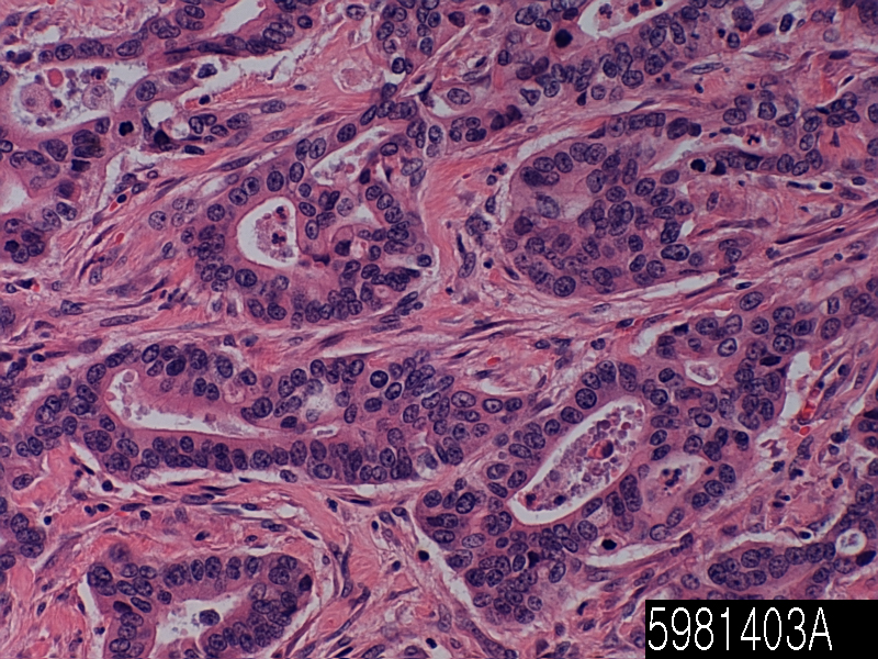
Cat. No. 5981403A
No. of samples 1
Description pancreas, adenocarcinoma
Age/Sex : 71/M
Price 197 EUR
260 USD
170 GBP

Pancreas adenocarcinoma

Product Related Literature

Pancreatic adenocarcinoma is a malignant tumor derived from a cell transformed has been made in pancreatic tissue. Generated in the exocrine pancreas component, the most common type of pancreatic cancer, which accounts for 95% of these tumors are (tumors showing a glandular architecture light microscopy) adenocarcinoma. The event is raised by the pancreatic islet cells, a small percentage can be classified as neuroendocrine tumors. Signs and symptoms, location of the tumor tissue Finally, based on the type and size, lead to diagnostics and can include jaundice abdominal pain, and back pain.

Pancreatic cancer is the most common cause of Fourth of cancer-related death in the United States and around the world eight. There is a very poor prognosis pancreatic cancer: all stages, total 1 – respectively, 5-year relative survival rate of 6% and 25%, local failure of the 5-year survival rate is the median survival for locally advanced , shows more than 80% of people together to be about 15%, in metastatic disease, is about 10 6 months. However, people are different – some, they are diagnosed only if they are terminally ill already, therefore, we have a few weeks or a few days. Even if you do not work, others will be able to have a slow progression, to live several years. Men, women and more the possibility of more than 30% higher in pancreatic cancer. Possibility of human origin and nationality of Africa will develop pancreatic cancer than white people is high.

It is debatable whether a risk factor for pancreatic cancer alcohol consumption. In general, the association constant is low, most of the research does not allow what the relationship. Compared chronic pancreatitis, and other types, in turn, often predisposition cancer associated with alcohol consumption, pancreatic cancer, chronic pancreatitis, the main cause too much alcohol chronic pancreatitis is not a precursor of pancreatic cancer . In some studies, the relationship between risk is shown to increase with alcohol intake. On the order of drink four or more, risk is the highest in heavy drinking per day in particular. Because it can not be a risk factor of “cancer, may make it to the alcohol level, it is an increased risk for people who consume alcohol to 30 grams per day about two spirits / day more and more people “The analysis pooled, concluded:” pancreas that there is no is not known. Our findings, risk of cancer of the pancreas and the consumption of 30 g or more alcohol per day is one and a slight increase in research or. “Some that I will warn that the findings of them, and there is a possibility of confounding factor is the cause. Even if there is a link, “may be due to the content of alcoholic drinks some” other than alcohol itself,. It was found that the risk is low to drink white wine in the Dutch study.

The majority of jaundice patient pain experience of pancreatic cancer, or weight loss,. Pain is present in 85% of 80% to patients with metastatic disease or advanced locally advanced. Typically, pain is felt in the dull pain and abdominal radiation on a cross back. Well, it is exacerbated by eating even intermittent. it is possible to weight loss is deep, it may be associated with anorexia, early satiety, diarrhea and steatorrhea. In most cases, jaundice is accompanied by itching and dark urine. Jaundice painful is present operable to be present in approximately half of painless jaundice patients and disease unresectable locally, in about half of patients with curable lesions potentially.

Initial performance may be dependent on the location of the cancer. Those of the head of the gland cancer and or tail usually present pancreas body is present steatorrhea, weight loss, jaundice in generally in weight loss and pain. Atypical diabetes, it is recent, but the recent emergence of the attack in front of pancreatitis history of thrombophlebitis or unexplained (Trousseau sign), has been pointed out frequently. Courvoisier is able to determine the gallbladder was painless jaundice extension showing strongly pancreatic cancer exists, signatures, and using the differentiation of pancreatic cancer by gallstones. Fatigue eat the pain, difficulty and irritability are also present. In most cases, pancreatic cancer was discovered during the evaluation of the above symptoms.

The liver function tests, may combination of results showing (raised level conjugated bilirubin, γ-guru Tamil transferase, of alkaline phosphatase) bile duct obstruction is displayed. The (carbohydrate antigen 19.9), in many cases, is a tumor marker that is increased in pancreatic cancer CA19-9. However, I lack the sensitivity and specificity. If you turn off on top of the 37 that have been using the U / ml, this marker, you will have a specificity of 87% in benign sophisticated from malignant disease and sensitivity of 77%. CA 19-9 can be can be normal at the beginning of course, increase as a cause of benign biliary obstruction. For example, an imaging test computer tomography and endoscopic ultrasound (CT), such as (EUS) may be used to identify the position and shape of cancer. Definitive diagnosis is made by surgical removal of tissue suspected radiological or endoscopic needle biopsy. In many cases, endoscopic ultrasound is used to guide the needle biopsy procedure visually. However, in general, pancreas, is arranged by using a scanner. In fact, histological diagnosis is not required to serve for determining ablation histological analysis and tumor should begin chemotherapy.

Common bile duct adenocarcinoma

Product name Common bile duct
Cat. No. 5881403A
No. of samples 1
Description common bile duct
Age/Sex : 54/F
Price 197 EUR
260 USD
170 GBP

Common bile duct adenocarcinoma

Product Related Literature

Bile duct is one of a long cylindrical structure carrying the bile. Bile required for digestion of food, intestinal secreted from the liver into the passageway carrying bile join hepatic duct to (carry bile from the gallbladder) and cystic duct to an opening in the inside, to form the common bile duct. Bile duct (see below) is a whole network of various sizes of the branch pipe in the liver. They are taken to be accumulated in the blood instead of active ingredient in the bile and gut (bilirubin), bile duct cancer blocked, gallstones, and scratches injury prevents the bile from there. This condition leads to jaundice, where the skin and eyes become yellow of bilirubin. The condition is this, I cause intense itching of bilirubin deposition in tissue. In some types of jaundice, chair would be much thinner than normal urine when it becomes too dark. Instead of being lost in the feces through ampoules Vater some, it is due to the all of bilirubin blood flow, which was filtered in the kidneys in the urine.

Jaundice, gall bladder can be caused not only patients rhinolith usually, pancreas, bile duct cancer, biliary tract cancer, and gallstones, leading to obstruction of the bile duct through the cancer part of the scar injury following, with conditions such as pancreatic cancer, such as the bile duct during the removal of. Doctors and, in general, by the intervention of the radiologist, biliary drainage is performed using (called biliary catheter or biliary stent, and bile duct leak) or a catheter tube. It can be used for biliary tract, such as surgery to relieve the blockage of a temporary solution or permanent treatment especially before. The drain can be inserted percutaneously through the liver, the procedure is called percutaneous transhepatic drainage and (PTBD) thereafter. Furthermore, then it can be carried and interventional radiology, as part of a percutaneous transhepatic cholangiography. It is diagnose or monitor a disease which can be used to take a sample of biliary bile drainage, as well as providing a route of administration of the drug substance.

It is a common form in the second (after the squamous cell carcinoma) of Eastern Europe, adenocarcinoma is the most common type of lung cancer in most U.S., Japan and Western Europe. Adenocarcinoma is extremely heterogeneous tumor that occurs (acinar, papillary bronchioles, solid) as well as a rare variant of some organization of the major four architectures. In most cases, however, these lesions were shown to be subdivided as follows: “China cancers. Different subtypes”, a mixture of variant or subtype of two or more, the many smokers, there as a case of lung cancer in the world, acinar tissue structure model is a histological subtype most common adenocarcinoma, which accounts for about 40% of adenocarcinoma far, the incidence increased It is decades of fairly recent you have. In Europe, adenocarcinoma will be able to include the architecture model dominant in 50-60% degree of adenocarcinoma.

Lung cancer is a disease very deadly. 5-year survival 22% and about 16%. Overall, survival was excellent in all phases (nipple) or bunch model of the patient as compared to patients with solid pattern rather but significantly worse than the bronchioloalveolar model. Survival rate was significantly better (ie, tube glands and / or have been developed more fully) than poorly differentiated when (simple gland ie) the cancer for patients with well-differentiated. In some studies, the Fez and cancer gene of H-Ras, to be an important driver of carcinogenesis of lung adenocarcinoma type of many has been shown.