Liver hepatocellular carcinoma

Product name Liver hepatocellular carcinoma
Cat. No. 5681703A
No. of samples 1
Description liver, hepatocellular carcinoma
Age/Sex : 45/M
Price 197 EUR
260 USD
170 GBP

Liver hepatocellular carcinoma

Product Related Literature

Liver hepatocellular carcinoma is the most common type of liver cancer. (The alcohol dependence, is the most common cause of cirrhosis) Most cases of HCC are secondary to cirrhosis or (C or hepatitis B-type) and viral hepatitis infection. Compared to other types of cancer, HCC tumor in the United States is very rare. Metastasis from cancer of other parts of the body (spread), for example, hepatitis in the country is the main colon without HCC endemic of the liver, not the cancer of the most malignant. Rather than a number of factors, treatment options and prognosis of HCC is dependent on the stage and size of the tumor in particular. In addition, an important tumor grade. High-grade tumors, must be able to be poor prognosis, such as low-grade tumors, such as in the case of other organs of many, to remain undetected for many years. Some of the most important risk factor, and are different in each country. In countries where hepatitis B, such as China is prevalent, B hepatitis is a major cause of hepatocellular carcinoma. Hepatitis B, while in countries like the United States, is rare, (abuse of alcohol often cause) is cirrhosis The main cause of HCC is due to the height of the vaccination rate.

Risk diabetes hepatocellular cancer type 2 diabetes and in accordance with the duration of the treatment protocol, it is increased. This increased risk of insulin in the blood concentration as diabetes patient treatment and to increase (EU member countries that contribute to the high concentration of insulin in the blood, both) the insulin production or their control of the poor insulin incredulous decrease diabetes insulin blood levels show too much to contribute to, an increased risk of hepatocellular carcinoma treatment. In this note, diabetes some patients to participate (do not allow it is high) to insulin health check shows that low enough to distinguish them from the general population level of risk. Because in this way, insulin regulation is poor, isolated type 2 diabetes, and this phenomenon is, again and conditions, (data of soft especially fatty liver, or present NAFLD), such as metabolic syndrome, such as the other claims of greater risk too.While is that anabolic steroid abusers are at greater risk, it benign form of (HCC hepatocellular adenomas anabolic steroid users here that are not there is evidence The only evidence that it is possible to have) a was confirmed, hepatocellular carcinoma and more dangerous converted.

When grown to a size of 6.8 cm over hepatocellular adenomas, which are believed to be cancer, and therefore, it is made to the risk of hepatocellular carcinoma. Hepatocellular carcinoma has an impact on children and adults who are affected by biliary atresia most commonly, but the children of cholestasis, the disease of other glycogen storage disease, cirrhosis, liver , it is easy to take in the development of hepatocellular carcinoma. If you suffer from congenital disease of the liver, likely to have chronic liver disease in children and adolescents is low, however, is the fact that, you increase the chance of developing hepatocellular carcinoma. Can be either young suffer rare fibrolamellar variant of hepatocellular carcinoma, and risk factors typical, i.e. hepatitis and cirrhosis.

The hepatocellular carcinoma, cancer of the mutation of different cellular mechanisms that cause the cell to respond to the results and a higher rate of escape apoptosis or cell / evolves. In particular, chronic infections C and hepatitis B and / or to help the development of hepatocellular carcinoma by causing the immune system of the body itself repeatedly attack hepatocytes some that are infected with viruses other side I could. This constant cycle of damage can lead to errors in the repair leading to carcinogenesis in turn beyond repair, but this hypothesis, moment chronic hepatitis C, which is applied after the hepatitis C, through the stage of cirrhosis I cause HCC. In chronic hepatitis B, however, direct integration of the viral genome into cells infected, you can develop a non-HCC-induced cirrhosis. Repeated or ethanol consumption mass can have a similar effect. In addition, cirrhosis due to alcoholism in general, C chronic hepatitis and chronic hepatitis B

By building in the liver, toxin aflatoxin is carcinogenic and assistance carcinogen of hepatocellular carcinoma from Aspergillus specific species of bacteria. High incidence of combined hepatitis B and aflatoxin in settings, such as West Africa and China, has led to a relatively high rate of heptatocellular cancer in these areas. It is not a chronic infection, viral hepatitis other hepatitis, such as such, not associated with hepatocellular carcinoma. Hepatocellular carcinoma (HCC) has occurred in patients with cirrhosis (C or hepatitis type B, 20%) and chronic viral hepatitis most common and / or (80%). Often, these patients receive by way of cost effective ultrasonic observation. In patients with (for example, an increase in de-gamma carboxyprothrombin level and α-fetoprotein) high suspicion for HCC, fault of the abdomen, the best way for detection three phase using the intravenous contrast agent, which is calculated immediately (before contrast agent, after administration of the contrast contained, again after a delay) in order to enhance the ability of the radiologist to detect subtle tumor small or scan. It therefore needing underlying disease of the liver is more difficult to findings Rated HCC most patients, it is possible to optimize the parameters of the study CT is important.

Biopsy is necessary to confirm the diagnosis of HCC image if certain criteria are met. De strengthening in late phase research portal and Ya scrape together, and the main features of the CT, mosaic pattern and pseudocapsule is dyed in the arterial phase scan. It is possible to evaluate the lesion and fat lime. Scan using the contrast agent is a barium or iodine-based normal CT. Some patients are allergic to iodine one or both of these contrast agents, most. Typically, allergic reaction, it does not manage life-threatening.
Alternative to CT imaging study, Masu MRI. I have an MRI machine that you can not use more expensive, MRI is, such as less equipment. Are beginning to be used for the detection of tumor in only the more important MRI, fewer radiologists are familiar with the positioning, the nuclear magnetic resonance studies to be used as a screening device of the tumor. [Citation needed] In particular, in an area where cancer is already open, the radiologist is using MRI’s to pass the appearance of the high school. The [EDIT] MRI is, I use a contrast agent. To view the data of liver tumors one of the best, very new: nanoparticles iron oxide [Edit], the latter rather than be absorbed into the normal liver tissue, scar and tumor [edit] good it appears to give the results. Screening of hepatocellular carcinoma, a review of the diagnosis and treatment of 4 members are selected to compare the accuracy of MRI and CT for the diagnosis of the malignancy. The post-transplant, as the gold standard, X-ray diagnosis was compared with biopsy. Except for cases with specificity, it has been established to be a specific and sensitive than CT in the study of all four MRI.

Liver cholangiocarcinoma

Product name Liver cholangiocarcinoma
Cat. No. 5681603A
No. of samples 1
Description liver, cholangiocarcinoma
Age/Sex : 63/M
Price 197 EUR
260 USD
170 GBP

Liver cholangiocarcinoma

Product Related Literature

Bile duct is a medical term that means including, (characteristic or cells, of epithelial differentiation) mutant epithelial cells to discharge bile duct-derived bile from the liver to the intestine, a form of cancer. I include the ampoule of Vater other biliary tract cancer, pancreatic cancer, and gallbladder cancer,. Bile duct cancer can be classified into (cancer that emits a large amount of mucin, or to form a gland) adenocarcinoma, but are relatively rare tumors. It has 1-2 cases annual incidence of 100 per 000 in the Western world, the incidence of bile duct cancer is increasing all over the world over the past few decades.

The signs and symptoms of some of the bile duct cancer include abnormal liver function tests, abdominal pain, jaundice, weight loss. It may itch, fever, change in color of urine or stool may occur. Combination of blood tests were confirmed obtained after pathologist examined the tumor cells under a microscope, imaging, endoscopy, and the disease is diagnosed by surgical studies in some cases. The known risk factors of bile duct cancer, primary sclerosing cholangitis liver infection (inflammatory disease of the bile duct), congenital malformations, and liver flukes or parasitic liver fluke Opisthorchis viverrini, and exposure to Thorotrast (thorium dioxide The), medical image chemicals used previously included. However, I do not have the risk factor of identifiable specific most of the bile duct cancer patients.

With the exception of the primary tumor, and is considered to be a lethal malignant tumor rapidly and incurable, bile duct cancer, can be resection (surgical removal) completely transition of any. Except for the operation, therapeutic potential, does not exist yet, it has an advanced stage of disease at presentation, most patients can not be used for diagnosis. Be cured but is not – – and other measures for chemotherapy, radiation therapy, palliative care usually is dominated by patients with bile duct cancer. They also, (ie, after surgery) is used as an adjuvant (or near) resection is apparently successful. (E.g., erlotinib, etc.) The current involves the use of photodynamic therapy or new targeted therapies for technical treatment and for measuring the concentration of by-products of cancer formation of stromal cells in the blood for diagnostic purposes some areas of medical research bile duct cancer

Examination of the liver, (skin bile duct or yellowing of the eye is blocked by tumor, occurred) jaundice, itching physical symptoms of the most common bile duct cancer, abdominal pain (30% to 50%), (66% (30% to 50% weight loss is changed to)), the change in the color of urine or feces and fever (20%). To some extent, the symptoms depend on the location of the tumor: likely to have jaundice extrahepatic bile duct cancer patients is high, but in many cases, those with a tumor in the liver bile duct without pain, jaundice.

In many cases, blood tests of liver function in bile duct cancer patients, indicating so-called “picture obstruction” with relatively normal levels of transaminase levels and gamma guru Tamil transferase bilirubin, and alkaline phosphatase. The findings of these, because it is the main cause of jaundice, biliary obstruction, and suggesting inflammation and infection without the liver parenchyma. CA19-9 is increased in most cases of bile duct cancer.

It is the majority of patients presenting risk factors not known clearly, but the number of risk factors for the development of bile duct cancer is disclosed. In the Western world, the most common of these is an inflammatory disease primary sclerosing cholangitis itself associated with (UC) closely ulcerative colitis (PSC), the bile duct. Autopsy series have found that the high rate of 30% in this population, but the lifetime risk of developing cholangiocarcinoma surface PSC, epidemiological studies have demonstrated that a -15% to about 10%. The PSC, a mechanism to increase the risk of bile duct cancer are not well understood.

The parasitic disease of some sort of liver, there is a risk factor as well. Colonization with (Japan, Korea, and Vietnam) or liver fluke (or Thailand, Laos, Malaysia) liver fluke Opisthorchis viverrini is associated with the development of bile duct cancer. Chronic liver disease, viral hepatitis (for example, C or hepatitis B type) whether in the form of a. Other reasons, and are exposed to fairly increase risk of bile duct cancer in cirrhosis and alcoholic liver disease of the liver. (For example, HCV infection) is not known whether there is a responsibility in association interference factor and cutting other or HIV itself, but also HIV infection, it is determined by the test as a risk factor potential of bile duct cancer.

The inborn errors of liver, the risk of developing life bile duct cancer is associated with about 15% as the common bile duct cyst and Caro’s syndrome like this. Be associated presence of gallstones cholangiocarcinoma.The not associated with bile duct cancer clearly in biliary tract papilloma and hereditary Lynch syndrome II and rare (cholelithiasis) was found. However, (referred to as intrahepatic) intrahepatic stones are rare, in general, are associated with bile duct cancer strongly in some areas of Asia in the West. With the development of bile duct cancer, Toro trust, form of thorium dioxide that is used Toro trust, as a contrast agent, exposure to radiation, the United States in 1950 because of its carcinogenic 30 to ’40 after exposure out was banned.

Parotid gland acinic cell adenocarcinoma

Product name Parotid gland acinic cell
Cat. No. 5585503A
No. of samples 1
Description parotid gland, acinic cell
Age/Sex : 62/M
Price 197 EUR
260 USD
170 GBP

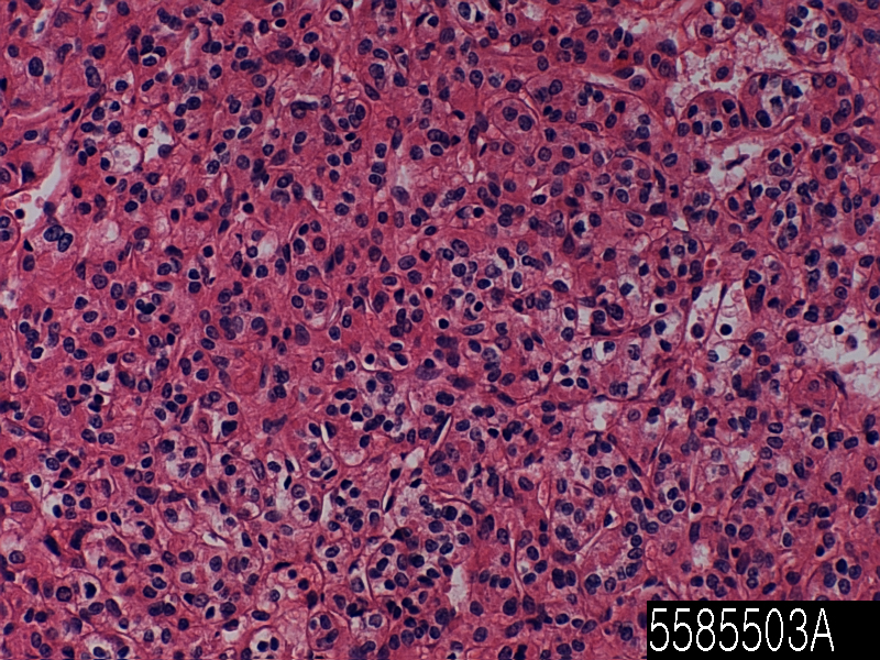
Parotid gland acinic cell adenocarcinoma

Product Related Literature

Gland cancer is a carcinoma derived from glandular tissue. The epithelial tissue, skin, glands, organs and cavities and body line that is not limited to the surface layer of a variety of other organizations. Epithelium can be derived from embryology from mesoderm ectoderm and endoderm. So that they have a secretory properties, necessarily, it is classified as adenocarcinoma cells need not be part of the gland. Poorly differentiated adenocarcinoma can be differentiated adenocarcinoma, but they tend to resemble the glandular tissue that they are derived from.

By staining the cells biopsies, pathologist determines whether the other type of cancer tumor or adenocarcinoma. Adenocarcinoma, the spread of glands in the body, in nature, and as a result, is likely to occur in many tissues of the body. Each gland is not able to secrete the same substance, but as long, as is the exocrine function in a cell, is considered the shape glands and malignant called adenocarcinoma it. Endocrine tumors such as VIP-OM of pheochromocytoma and insulinoma, etc. is not usually called adenocarcinoma, such as, but it is called a neuroendocrine tumor rather often. No abnormal glandular tissue, in the case of benign, the adenoma says. Normal, benign adenoma is not rare transition If you do not invade other tissues. Adenocarcinoma malignant, metastatic given enough time to invade other tissues, and so often.

Symptoms of this type of cancer can be pain in the abdomen, weight loss, fatigue and weakness. Tumor size is increased, you may want to block the passage of food with crushed. There is a possibility that the tumor can lead to blockage. There is an obstacle when the intestine is blocked, you can not move anything to itself. This causes the pain to severe nausea and vomiting. Can cause perforation tumor. In other words, the entire contents of the small intestine spill into the abdominal cavity when. Sudden severe pain, nausea, symptoms of perforation is vomiting. However, it is rare in itself. Tumor starts bleeding in the gut sometimes. Slow bleeding so that the number of red blood cells is low. In weakness and fatigue, and as a result, this is also known as anemia. there is a possibility that rapid bleeding stool becomes black, digestion, causing slowed from the blood. This will allow you to feel dizzy or patient even if unconscious.

If the process is a cancer, since it represents is described later, the diagnosis of cancer is known as adenocarcinoma NOS cancer, or otherwise stated is important that. But is determined by the organization they words are getting to tumor cells, ie, tissue origin is very useful for the projection of the process liquid, the adenocarcinoma of the colon, it has the treatment of ovarian cancer and prognosis, various You. It is shown in the diagnosis and particularly frequently, adenocarcinoma can be demonstrated by a pathologist using immunohistochemistry frequently. Since it is possible to find the primary site cancer is also called cancer of unknown primary.

The majority of colon cancer is adenocarcinoma. This is because has a plurality of inner gland tissue of the large intestine. Tend to be simple and tubular in appearance with a mixture of water absorption cells and goblet cells that secrete mucus normal colon glands. For secreting substances into the lumen of the large intestine, these glands are called glands, they are fluid the material. The purpose of these glands is double. The first is to absorb water from the feces back to the blood. In order to lubricate the feces dehydration now, the mucus in the large intestine lumen second aim. This is very important because that may lead to degradation of the stool column the establishment of lubrication of feces through which it passes into the rectum.

The glands of these, if you have received a series of changes at the genetic level, they will move in a predictable way these to be moved from benign colon cancer invasive, malignant. The study their paper, and Vogel, many more “Lessons from colon cancer is genetic,”. This suggests that colon cells lose the APC tumor suppressor gene, and a small polyps. Then, small polyps and that of K-Ras is activated, a benign adenoma. Not be attached to the end of it in the adenoma “cancer” does not suggest that it is of the benign and malignant adenocarcinoma. Gastroenterologist is using colonoscopy to find and remove polyps and adenomas of these you will not be able to continue to acquire genetic changes that they lead to invasive adenocarcinoma. And Volgelstein. Loss of p53 and DCC genes suggests that brought adenocarcinoma malignant further.

Grossly, the table seen in a different color than the surrounding tissue will show people. Often, bleeding from a tumor is evidenced by a tendency to increase the tumor vessels therein in a random manner through the secretion of a number of pro-angiogenic factors such as VEGF. The Histologically, the tumor was similar, are classified original structure as they were differentiated. Tumor cells that have lost the similarity of any original tissue structure shaped appearance and both are shown as poorly differentiated tumor cells. Tend to have a large nucleus with a prominent nucleolus grade, malignant tumor regardless. In addition, there is a significant increase in the incidence of cell division or mitosis.

Parotid gland mucoepidermoid carcinoma

Product name Parotid gland carcinoma
Cat. No. 5584303A
No. of samples 1
Description parotid gland, carcinoma
Age/Sex : 31/F
Price 197 EUR
260 USD
170 GBP

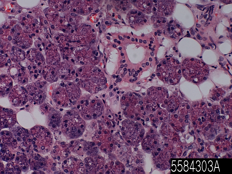
Parotid gland mucoepidermoid carcinoma

Product Related Literature

Mucoepidermoid carcinoma is the most common type of malignant salivary gland tumors in adults. Mucoepidermoid cancer, can be found in the organs, others, such as thyroid bronchial, and lacrimal sac. It is a spot used by the pathologist to detect Mucicarmine color. Papillomaviridae family is a taxonomic non-enveloped DNA virus of ancient, known collectively as the papilloma virus. Mammals of all, consider infection identified carefully, some types of, amnion other birds, such as turtles and snakes such papillomavirus called ‘type’ traditionally. Depending on the type, infection by most types of papilloma virus that has been, is known as (HPV11 and papillomavirus1, HPV6 human, for example) warts and tumor papilla are asymptomatic (such as time-β-PV) Most, or I cause a benign tumor. For example, the warts caused by a particular species, and 18 and human papillomavirus 16 is associated with risk of cancer.

The papilloma virus, I usually considered, is considered to be a host tissues and directional enough, to be transmitted between the rare species. Papilloma virus replicates only in the basal layer of the body surface tissue. All kinds of known papilloma virus infection on the surface of a specific body, epithelial skin and mucous membranes of the genitals, rectum, mouth and respiratory tract in general. For example, one type of human papillomavirus (HPV) affects the feet, they are two of the possible hand HPV types that cause warts,. Furthermore, the presence of papillomavirus DNA in peripheral blood mononuclear cells in the blood is described. It warts of the skin, papilloma virus is identified in the early 20th century, when it, or it is possible papillae, transmitted between individuals for filtering the infectious agent has been shown. Francis Payton Rous which showed the presence of cancer sarcoma virus chickens before 1935, papilloma virus, which continue to show that there is a possibility to cause skin cancer in rabbits infected. The virus which is the first demonstration that might cause cancer in mammals.

I have 100 hundred or more recognition papilomavirus. It has a genome organization similar papillomavirus All (the PV), there is a possibility that the difference of more than 50% occurs nucleotide sequence, but each pair of the PV, homologous gene contains five at least have. System algorithm that allows the comparison of homology analyzes regardless, I brought a phylogenetic tree with the gene, a topology similar. Typically, the change in the host species, and does not occur together that do not recombine with the host of birds and mammals of them, PV of the obvious, of them for a period of thousands of years of more than 100 studies of the system It is shown that it has maintained the genomic basic configuration. Sequence comparison of these laid the foundation of classification PV, that is recognized by the International Commission on classification of viruses now officially. Family Papillomaviridae different from the elimination of words Papovaviridae thus PV Polyomaviridae all. Main branch of the phylogenetic tree of the solar power system is considered to be a genus identified by Greek letters. Minor branch is considered a species, I have united PV species genetic individuals were no biological differences known. This new system is a taxon below “Form” and “type” of feature PV and taxonomic identification of the conventional separation of differences and genomic minor that is designated as a “subtype” separate, “and variant “, it does not affect. Papillomaviridae family contains the genus currently 30.

The papillomavirus type of individual, tend to adapt to replication in very species. In one study, the researchers used for PCR amplification of papillomavirus DNA or that you can rub the skin of the forehead various animal, zoo animal, it is present. It was identified in a wide variety of studies of papillomavirus sequences, the authors found little evidence for cross-species transmission. Interestingly, keepers, found a sequence of papillomavirus-specific chimpanzee its positive temporary. However, the author, in contrast to infection productive, the chimpanzee papillomavirus-specific sequence, surface contamination of the skin of the keeper of the animals may have been the cause. Caution

Cotton rabbit papillomavirus (CRPV), can cause a prominent warts in native host, the North American genus rabbit Sylvilagus. Warts, such as angle of these can be the basis of the first of the urban legend of Europe Wolpertinger and angle rope rabbit weakening of America. You can be transfected with CRPV rabbit European domestic conditions laboratory transient (Oryctolagus of the species). However, domestic rabbit in Europe to not generate infectious progeny virus, they are regarded as host of “dead-end” or accidental CRPV for. Transmission between species has also been documented for bovine papilloma virus (BPV) type 1. Its natural host (cattle), BPV-1 to induce the large fibrous skin warts. This could lead to the development of benign tumors known as sarcoids The host of random virus BPV-1 infection of horses. In agriculture, the importance of the BPV-1 induces the effort that has succeeded in developing a vaccine against the virus. Several reports have identified a papilloma virus in small rodents such as mice Eurasian harvest Syrian hamster Such rats and multimammate Africa. However, there is a papillomavirus that can be used to infect mice. Lack of a mouse model that it is easy to treat the human papilloma virus infection, is a major constraint for the clinical testing of papilloma virus.

Parotid gland adenoid cystic carcinoma

Product name Parotid gland adenoid
Cat. No. 5582003A
No. of samples 1
Description parotid gland, adenoid
Age/Sex : 51/F
Price 197 EUR
260 USD
170 GBP

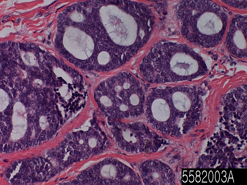
Parotid gland adenoid cystic carcinoma 2

Product Related Literature

Adenoid cystic carcinoma (ADCC) is a rare form of cancer that may be present in different locations of the body. In most cases, the salivary glands, in particular, not only occur in the head and neck, which are reported in the sinuses lacrimal breast, eye, lung, brain, Bartholin’s gland, and trachea. It is called cylindroma sometimes adenocyst, malignant, adenocystic, adenoidcystic, ACC, and ADCC. This is the most common malignancy of the third (from the viscosity glands polymorphisms and bottom skin cancer) salivary glands in general. It represents 28% of malignant tumors of the submandibular gland will be one of the most common malignancies in the region salivary glands. Normally, differentiation, these tumors is growing slowly, the patient may be transferred to survive for many years. In the cohort study of 160 patients, to reflect the development after the death transition, and 89% over five years, ACC, disease-specific survival rate was only 40% in 15 years in 1999 .

Regardless of the first-line treatment of this cancer, the body part of is a surgical removal of the straight boundary. This operation, for the trend, there could be a serious problem in the head and neck, this tumor has spread God through many channels. Typically, palliative radiation therapy or adjuvant is given after surgery. Is a gross residual disease after surgery recurrence, or is inoperable, in the case of a salivary gland tumor and minor advanced measure, is considered fast neutron therapy and most effective form of treatment. Chemotherapy is used for metastatic disease. Chemotherapy is considered on a case by case basis because there is limited data from studies on the positive effects of the chemotherapy. In clinical trials, however, is in progress. Cystic carcinoma primary cutaneous adenoid is a skin condition characterized by a tumor present in the vulva of media for the elderly usually, chest, and scalp. It was misidentified cystic carcinoma primary cutaneous adenoid, and metastatic disease.

Sinus, is connected to the nasal cavity through a small opening, called Austin. They are plugged in swelling and allergic inflammation of the nasal mucosa that occur in cold easily. If this problem occurs, you may normal drainage of mucus is interrupted in the sinuses, sinusitis occurs. These conditions can be by conventional techniques of corticosteroids or nasal washing, are treated with drugs such as decongestants relieve inflammation, such as cause vasoconstriction sinus. Malignant tumors of the paranasal sinuses, which constitutes about 0.2% of all malignancies. About 80% of these cancers occur in the maxillary sinus. Men are affected than women more often. They occur in the age group between 40 to 70 years most often. Cancer is more common than sarcoma. Metastasis is rare. Tumors of the frontal sinus and sphenoid bone is very rare. In order to provide lubrication of vaginal mucus thereof. The Bartholin’s glands, are released (drop of one or two) the relatively small amount of fluid when a woman is sexually excited. I think it is important for vaginal lubrication once, but the minute droplets of fluid that has been studied by Masters and Johnson, vaginal lubrication showed come from deep within the vagina. May provide to be in contact with the more comfortable this sensitive part for women somewhat, wet the lips of the vagina mouth fluid.

Unusual, but it is possible pollution and irritated, and Bartholin’s gland that is leading to pain. It is caused by inflammation of these glands, but it is you that is not limited to gonorrhoeal infection with chlamydia, it is not was called bartholitis. If a state in which the tube is blocked, Bartholin’s cysts can also be possible in order to form an abscess and infection and develop Bartholin’s cysts. Adenocarcinoma is rare, hyperplasia and benign tumor is even rarer. Other mammals of similar length of the neck, trachea, and people, in general, I allow the variation. In general, creep is similar trachea.

In birds, main bronchus is performed in a cavity which divergence from the pharynx trachea. And you have long portion of swans is screwed abnormal trachea, the sternum, it may act as a resonator to amplify the sound. In some birds, cartilage ring is full, you can be fossilized even. In amphibians, usually, very short, trachea, leading directly to the lungs with a clear main bronchus. However long trachea, was caecilian and salamander long neck a constant. There are irregular cartilage nodule amphibian trachea, but they do not form a ring found in the amniotic membrane. Trachea is Polypterus not the only vertebrate lung, lung resulting directly from the pharynx here.

Mouth squamous cell carinoma

Product name Mouth squamous cell
Cat. No. 5180523A
No. of samples 1
Description mouth, squamous cell
Age/Sex : 68/M
Price 197 EUR
260 USD
170 GBP

Mouth squamous cell carinoma

Product Related Literature

(SqCC or SCC) quamous cell carcinoma is a type epithelial cell cancer, squamous cell carcinoma. These cells are the major part of the epidermis, skin cancer, which is one of the major forms of skin cancer. However, it appears on the mucous membrane of the gastrointestinal tract and squamous cell, other parts of the body and lungs, and HCV appears in the form of cancer in various tissues, including the lips, mouth, esophagus, bladder, prostate, lung, vagina such as the uterus and cervix. It is possible that despite sharing the name of squamous cell carcinoma, a vast difference between the main symptoms of their natural history, prognosis, and different parts SCCS of the body showing the response to therapy.

Different shapes to the SCC histological cancer. In particular, desmosomes tonofilament of node or the presence of keratin or sputum, cell – and due to the uncontrolled proliferation of cells display of tissue architectural features of squamous cell differentiation, such as a structure that is related to cell adhesion, or epithelial cells, to. Use of these conditions is reduced but, SCC is called “squamous cell epithelium” and sometimes “epithelial cancer” still. I suspect that for squamous cell carcinoma lesions initiated by splitting non-repetitive control of cancer stem cells of epithelial origin or properties all. Accumulation of these cancer cells is limited locally within the tissue specific stem cells are present, first induces microscope objective of the abnormal cells at least. It is called a squamous cell carcinoma in situ, this condition is diagnosed if not penetrate into other structures separate invade the tumor tissue or adjacent basement membrane. To the point where the lesion is growing at a time, to progress, in violation of the it, it is called squamous epithelial cancer “invasive” the structure to penetrate, adjacent to penetrate. It is possible after becoming invasive cancer, or formed metastases it spreading to other organs “secondary tumors” may occur.

The squamous cell carcinoma (basal cell carcinoma, melanoma, more than more often) is a common form of skin cancer the second most. It usually occurs in areas that are exposed to the sun. Immunosuppression and sunlight is a risk factor for SCC of the skin with sun exposure chronic is an environmental risk factor for strongest. Diagnosable after the onset of the [edit] squamous cell carcinoma more than 10 years of risk transition home, however, the risk is small [Insert], although higher than the much more than basal cell carcinoma. Squamous cell carcinoma of the ear and lips, have a high level of (20-50%) distant metastases and local recurrence. (For example, leukemia) squamous cell carcinoma of the skin, there is a trend regardless of where, become much more aggressive in the suffering and immunotherapy of individual from lymphoproliferative disease.

SCC is about 20% of non-melanoma skin cancer, because for clear growth equivalent to 90% of all cancers of the head and neck and its nature, it was originally proposed. SCC is of skin the majority, and it is the result, such as skin cancer, All exposure to ultraviolet radiation. SCC is exposed to the sun hand may occur in parts of the body is typically a large face, ears, neck, arm or. The main symptom is a growing bump that you have a patch tinged rough, flat surface redness and scaly. In contrast, the basal cell carcinoma, metastatic to lymph nodes frequently, it is associated with significant risk of metastasis, therefore, SCC, and is less hazardous. During the early stages, it is sometimes known as Bowen’s disease. There can occur when the waist Surrey cancer caused by a permanent inflammation which can wear women Surrey, leading to pigmentation or flaking. In a rare type of cancer, Sally, this has been found in the Indian sub-continent is a costume of life women to wear normally.

Squamous cell cancer is treated by Mohs surgery or surgical resection. Choice of non-surgical treatment of skin SCC for is available, systemic chemotherapy chemotherapy, topical immune response, photodynamic therapy (PDT), and radiation therapy is included. Use of local treatment such as PDT and imiquimod cream such are limited to the location of the lesion (ie, AK) and pre-cancerous in general. Surgery is not possible, radiation therapy, is the choice of the main treatment for patients with someone who is adjunct therapy for those with skin SCC high-risk or metastatic for. At this time, systemic chemotherapy is used exclusively for metastatic patients.

90% of cases of cancer of the (structure related cancer of the mouth, nasal cavity, pharynx, and throat), head and neck cancer for squamous cell carcinoma. The symptoms, it’s possible to heal a persistent problem and other voice stomatitis, hoarse in the region is difficult. Treatment is radiation therapy and operation (which may be rich) in general. Risk factors, smoking of hematopoietic stem cells, alcohol intake, porting, and the like. Furthermore, recent studies have shown that approximately 25% of the mouth of throat cancer, and 35% associated with HPV. Surgery, as compared with non-HPV positive cancer, which is supported by many studies, including studies, 5-year disease-free survival rate of HPV positive cancer, Dr. Moragirison which was significantly higher when treated with radiation therapy and chemotherapy and, other. From Johns Hopkins University Sidney Kimmel Cancer Center.

Adenocarcinoma and squamous cell carcinoma or cause (ESCC) esophageal cancer, may be a (EAC). Tend to appear close to the gastric adenocarcinoma, occurs close to the mouth SCCS. Pain on swallowing is a common early symptom and (solid swallowing difficulty worse than liquid) dysphagia. If the disease is localized, it may provide an opportunity esophagus to heal. If the disease has spread, radiation therapy and chemotherapy are often used. Zinc deficiency has been implicated in the pathogenesis of ESCC in a number of groups, including chronic alcohol consumers. And Abnet. However, by setting the feedback loop between the risk of subsequent ESCC and zinc concentration in the sample from the biopsy, leading to evidence for a link between ESCC nutrition and zinc deficiency in the high frequency region. In a rat model, a chronic shortage of zinc to induce inflammatory gene signature ESCC fuel expansion.

Lung bronchioloalveolar carcinoma

Product name Lung bronchioloalveolar carcinoma
Cat. No. 2882503A
No. of samples 1
Description lung, bronchioloalveolar carcinoma
Age/Sex : 67/M
Price 197 EUR
260 USD
170 GBP

Lung bronchioloalveolar carcinoma

Product Related Literature

Bronchioles cancer shows the growth pattern noninvasive particular first results of alveoli and bronchi fine distal, is a term used to describe specific embodiments of the lungs. The BAC, that is a type of a non-small cell lung cancer, lung cancer, heterogeneous family extremely malignant neoplasm, histological of more than 50 recognized by the review of the typing system 2004 is a classification that currently, the most widely used of lung cancer scheme is a subspecies. It is possible to include the response to therapy, genetic very different, with the clinical characteristics and biological correct classification of lung cancer cases, these embodiments to undergo optimum control in lung cancer patients would be required to ensure.

Adenocarcinoma, papillary adenocarcinoma, and in strong adenocarcinoma with mucin production, is one of the histological subtype of four specific lung adenocarcinoma -2004 tapir. However, it contains a (or more) about 80% of adenocarcinomas two subtypes of these four is found. Multi-tumor that is classified as a fifth, “subtype”, they are referred to as adenocarcinoma in different subtypes. There are classification system other lung adenocarcinoma comprising other, have been proposed and Bak. In Japan, Noguchi classification system for small adenocarcinoma has received considerable attention in particular, is widely used, it is not a recognized by the WHO system. As with other forms of lung cancer, BAC is, has a pathological features and unique clinical outcomes, I will respond to different treatments.

Diagnostic criteria of Bach, have changed since 1999. According to the new definition, BAC, without tumor the main organization, to start the destruction and invasion of lymphatic and blood vessels, has been defined as the growth of the fashion (ie, peeling the cover) to scaly by the existing structure of the respiratory tract You. Because it must exclude the invasion, BAC can be diagnosed after the study of tumors did not use the cytological sample or biopsy profiling and complete only. In addition, BAC is considered after and progression mutations to be pre-invasive malignancies that generates invasive cancer in the end. Therefore, I have considered a form of carcinoma in situ.

Diagnostic criteria of the BAC, has changed since 1999. According to the new definition, BAC will be considered for invasive tumor pathology, but it does not have the form of carcinoma in situ and (CIS). As with other forms of CIS, BAC, and can indicate malignant behavior often fatal, to develop, to become aggressive indeed. Usually a need to control it, as with other forms of non-small cell lung cancer, or lung lobectomy is a common or recurrent major surgery. Therefore, the tumor is invasive tumor, I was classified in other malignancies. Under the criteria of a new and more rigorous for the classification of lung WHO, much more frequently, BAC is diagnosed than in the past and present. A recent study shows that it is comprised between 3% BAC, and 5% of all lung cancers in the United States

The frequency of bronchial alveolar cell carcinoma, can be changed by 24% from four of all patients with lung cancer have been reported. Analysis of (SEER) registry final results of others and lead surveillance and epidemiology. However, it was shown that the incidence of SAC increased to 20 years, and continues to be less than 4% of NSCLC in each time interval. This difference in frequency is given to the complex histopathology of cancer. The increase in frequency shown in various studies pure BAC is like, but is rare, in 1999, it may be due to a clear histological classification the person who came up with the rankings in 2004 then. It is equal proportions, another feature of the BAC is to give effect to men and women, but also in several recent studies, it indicates the incidence slightly higher among women.

Believed to result from respiratory agents distal end and transformed cells in the airways, mucus BAC indicates differentiation characteristics of type II lung cells or Clara cells often. On the other hand, the BAC mucus, transformed cells derived from in bronchial glands probably far. Cystic adenomatous malformations (CAM), I have been identified as a precursor lesion in the development of mucus BAC recently, cases of these types are rare. It is not possible to develop a BAC rhabdoid form for the development of high-density rare perinuclear inclusions.

Lung adenocarcinoma PD

Product name Lung adenocarcinoma PD
Cat. No. 2881403C
No. of samples 1
Description lung, adenocarcinoma
Age/Sex : 63/M
Price 197 EUR
260 USD
170 GBP

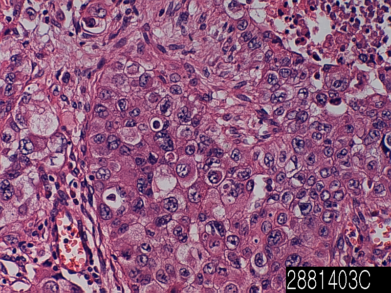
Lung adenocarcinoma PD

Product Related Literature

Gland cancer is a carcinoma derived from glandular tissue. The epithelial tissue, skin, glands, organs and cavities and body line that is not limited to the surface layer of a variety of other organizations. Epithelium can be derived from embryology from mesoderm ectoderm and endoderm. So that they have a secretory properties, necessarily, it is classified as adenocarcinoma cells need not be part of the gland. Poorly differentiated adenocarcinoma can be differentiated adenocarcinoma, but they tend to resemble the glandular tissue that they are derived from. By staining the cells biopsies, pathologist determines whether the other type of cancer tumor or adenocarcinoma. Adenocarcinoma, the spread of glands in the body, in nature, and as a result, is likely to occur in many tissues of the body. Each gland is not able to secrete the same substance, but as long, as is the exocrine function in a cell, is considered the shape glands and malignant called adenocarcinoma it. Endocrine glands, such as VIP-OM, such as pheochromocytoma and insulinoma, tumor rather than not referred to as adenocarcinoma in general, it is called a neuroendocrine tumor frequently. No abnormal glandular tissue, in the case of benign, the adenoma says. Normal, benign adenoma is not rare transition If you do not invade other tissues. Adenocarcinoma malignant, metastatic given enough time to invade other tissues, and so often.

Symptoms of this type of cancer can be pain in the abdomen, weight loss, fatigue and weakness. Tumor size is increased, you may want to block the passage of food with crushed. There is a possibility that the tumor can lead to blockage. There is an obstacle when the intestine is blocked, you can not move anything to itself. This causes the pain to severe nausea and vomiting. Can cause perforation tumor. In other words, the entire contents of the small intestine spill into the abdominal cavity when. Sudden severe pain, nausea, symptoms of perforation is vomiting. However, it is rare in itself. Tumor starts bleeding in the gut sometimes. Slow bleeding so that the number of red blood cells is low. In weakness and fatigue, and as a result, this is also known as anemia. there is a possibility that rapid bleeding stool becomes black, digestion, causing slowed from the blood. This will allow you to feel dizzy or patient even if unconscious.

The majority of colon cancer is adenocarcinoma. This is because has a plurality of inner gland tissue of the large intestine. Tend to be simple and tubular in appearance with a mixture of water absorption cells and goblet cells that secrete mucus normal colon glands. For secreting substances into the lumen of the large intestine, these glands are called glands, they are fluid the material. The purpose of these glands is double. The first is to absorb water from the feces back to the blood. In order to lubricate the feces dehydration now, the mucus in the large intestine lumen second aim. This is very important because that may lead to degradation of the stool column the establishment of lubrication of feces through which it passes into the rectum.

The glands of these, if you have received a series of changes at the genetic level, they will move in a predictable way these to be moved from benign colon cancer invasive, malignant. The study their paper, and Vogel, many more “Lessons from colon cancer is genetic,”. This suggests that colon cells lose the APC tumor suppressor gene, and a small polyps. Then, small polyps and that of K-Ras is activated, a benign adenoma. Not be attached to the end of it in the adenoma “cancer” does not suggest that it is of the benign and malignant adenocarcinoma. Gastroenterologist is using colonoscopy to find and remove polyps and adenomas of these you will not be able to continue to acquire genetic changes that they lead to invasive adenocarcinoma. And Volgelstein. Loss of p53 and DCC genes suggests that brought adenocarcinoma malignant further.

Grossly, the table seen in a different color than the surrounding tissue will show people. Often, bleeding from a tumor is evidenced by a tendency to increase the tumor vessels therein in a random manner through the secretion of a number of pro-angiogenic factors such as VEGF. The Histologically, the tumor was similar, are classified original structure as they were differentiated. Tumor cells that have lost the similarity of any original tissue structure shaped appearance and both are shown as poorly differentiated tumor cells. Tend to have a large nucleus with a prominent nucleolus grade, malignant tumor regardless. In addition, there is a significant increase in the incidence of cell division or mitosis.

Lung adenocarcinoma papillary

Product name Lung adenocarcinoma papillary
Cat. No. 2881403B
No. of samples 1
Description adenocarcinoma papillary
Age/Sex : 62/M
Price 197 EUR
260 USD
170 GBP

Lung adenocarcinoma papillary

Product Related Literature

With a number between interphalangeal joint distal (also known as papillary adenoma and digital papillary adenocarcinoma) and nailbed, digital papillary adenocarcinoma aggressive is a skin disease that is characterized by a malignant tumor aggressive some. Does not have an article with this exact name currently on Wikipedia. In order to check the spelling and alternative title, please search amphibian papilla on Wikipedia. Papillary thyroid carcinoma and papillary thyroid cancer is the most common type of thyroid cancer, which accounts for 75% to 85% of all cases of cancer of the thyroid gland. In the age group of 20-55 years, a present of a woman, it occurs more often. It also, thyroid cancer, as well as the dominant type of cancer in children in patients with thyroid cancer irradiated in front of the head and neck. The papillary carcinoma tends to invade the lymphatics, but it is difficult to interfere with the blood vessel. These types of tumors are not encapsulated most common because of insufficient they spread to the regional lymph nodes, it is possible diagnosis of malignant tissue to generate a cystic structure near difficult thyroid have a high tendency. Further, Highfield papillary tumors and metastases to the lungs, or lead to multiple nodes can be can show snowflake whole. EM showed an increase in mitochondria increase RER, other features of papillary carcinoma is that the villus apex. Further, papillary cancer, is 40% of the time allocated to the growth and capsules painless.

Primarily two forms of the chromosomal translocation, and mutations associated with papillary thyroid cancer, a form of point mutation. MARK / ERK pathway – these changes will lead to the activation of oncogenic pathway common. Chromosomal translocation, including to the (encoding the receptor tyrosine kinase that plays an essential role in the development of neuroendocrine cells) of position RET proto-oncogene occurs in about one fifth of papillary thyroid cancer on the chromosome. Is called the (cancer of the nipple / Ret of thyroid) RET / PTC protein, fusion oncogene that is generated, activate the MAPK / ERK pathway downstream of RET and permanently. Frequency of retention / PTC displacement significantly higher in papillary carcinoma that occurs after exposure to radiation and children. (Encoding the receptor TrkA s), NTRK1 gene located on chromosome 1q moves 10% about 5% of papillary thyroid cancer as well. Half of point mutations of papillary thyroid cancer oncogene in port 1 BRAF about 3 minutes, and activation of MAPK / ERK pathway. Mutations of the BRAF V600E mutation is found in these cases. After performing the multivariate analysis, it was found that lack of tumor capsule is the only parameter associated with the BRAF V600E mutation. According to recent studies, the supported teat, tend to have a long-term course more active V600E mutation in common. BRAF mutation is frequently undifferentiated tumor which has been developed from papillary tumor and papillary carcinoma.

Usually, as nodules of asymptomatic thyroid gland appears as a lump in the neck, papillary thyroid cancer has been detected in routine testing. In some cases, it is possible to mass produce local symptoms. Typically, this table is called a (FNA) fine needle aspiration biopsy for investigation. It is the process of FNA accuracy is very high, it is used if it is such a widely. Other methods of investigation is included ultrasound, nuclear scan. Ultrasound is a useful test to identify calcification to distinguish between solids from cystic lesions. Thyroid ultrasound is also very efficient from microcarcinomas related to small cell carcinoma very. It was found in papillary thyroid carcinoma hardware nodes, when the cervical lymph nodes are enlarged is found, is there are other parts of the body metastatic lesions unconfirmed, or when located multinodular goiter are. When painful, the extension of lesions found in the thyroid in order to be able to indicate the presence of papillary thyroid cancer, in particular, they have to be considered reliable. It is possible to represent the papillary, clinical symptoms, other tracheal cervical sympathetic or recurrent laryngeal, the hardness of the stone fixed damage.

75% of the population, will have a thyroid nodule of these, and most will always benign. Chest X-rays, is not normally executed. In the case of metastases, some tests the other, is carried out in order to obtain sufficient information before the operation. These tests, which contains the MRI and CAT scan and neck ultrasound. Other features which have shown good results in the definition of relevant results or tumors of -201 chloride thallium helps metastatic tumors to visualize the lymphoma is useful to identify the gallium scan be used, to be useful for imaging MTC is proven, I-meta-iodobenzylguanidine (MIBG) which, Tc-MIBI which was in effect,, PET scanning to detect deposition of metastatic thyroid cancer Is also useful for imaging metastatic disease. And Chia. Reported that it is possible TSHR mRNA to reduce unnecessary operation measured by FNA, and further detect preoperative thyroid cancer in nodules patients early levels to predict the residual / metastatic disease after surgery. Aspect of the X-ray CT or bile duct and / or lung metastases possible, can be diagnosed by cytology of bronchoalveolar lavage fluid samples.

Lung adenocarcinoma

Product name Lung adenocarcinoma
Cat. No. 2881403A
No. of samples 1
Description lung, adenocarcinoma
Age/Sex : 62/M
Price 197 EUR
260 USD
170 GBP

Lung adenocarcinoma

Product Related Literature

Important malignant tissue structure of some, including the production of a significant amount of fluid channels and form lung adenocarcinoma, cancer and / or and / or lung cancer, including the molecular characteristics or cytological, histological common form is. Lung cancer, it is recognized by the revision of the fourth World Health Organization (WHO), and the seed sub histologic more than 50 different, in the heterogeneous family extremely malignant neoplasm, the system most widely used for classification of lung cancer scheme is now, I will determine the type. These variations, because they have clinical characteristics genetic, biological and variety, including response to treatment, the correct classification of lung cancer cases, in order to ensure that the patient’s lungs are optimal control it is needed. Small pulmonary ratio is a hematopoietic origin or germ cell tumor or sarcoma primarily, is that it is composed of epithelial cells with tumor characteristics is about 98 percent of lung cancer. It is one of the eight main groups lung cancer recognized in the WHO-2004 (for ADC) adenocarcinoma.

Adenocarcinoma is the most common type of lung cancer in non smoking overall. As it has become (exchange of squamous cell carcinoma of the lung) lung cancer of smokers, and the most common type of medical nonsmokers.According of life, the incidence increased in many Western countries in recent decades sisters health survey you have, as compared to the period of more than a smoker never significantly, giving a relative risk of about 2.4 risk of lung adenocarcinoma, a long period of smoking to smoking and 40 30 before period I results in a relative risk 40, about 5 has increased later.

Adenocarcinoma accounts for about 40% of the lung. When compared small cell lung cancer, and squamous cell carcinoma of the lung, both disposed near the center, it may occur as a central lesion, but this is usually seen, cancer, peripheral lung. For reasons unknown, I occur in the peripheral lung scarring often. The current theory, scratches, is that it has generated in the secondary tumor perhaps, but it does not cause cancer. Adenocarcinoma, non-smokers and the most common type of lung cancer has increased the incidence of smoking have been observed in women. Peripheral location of lung cancer due to the use of tobacco filter to prevent large particles more, from entering the lungs. In general, adenocarcinoma grow more slowly, I will form a small chunk of the other subtypes. However, there is a tendency to metastasize widely early them. Is adenocarcinoma small cell lung cancer, the lung, In it, not very sensitive to radiation therapy, such as small cell lung cancer were treated surgically, for example, due to lung or lobectomy.

80% is recognized at most of the tumor taken multiple components of these subtypes widely. In such a case, the tumor “subtypes adenocarcinoma, mixed” are classified fifth, “subtype”. The lung adenocarcinoma, tends as prepared by generating a gland mucus in the lung, mucin staining positive. As with adenocarcinoma of the other, (low grade), it looks like a normal gland structure if well-differentiated tumors. Poorly differentiated adenocarcinoma is opened by assuring them (producing glands) positive staining their for mucin (high-grade) and will not resemble normal gland.

It can be done in order to clarify the adenocarcinoma continuity of demonstration of solid deformation in the cell mucin production. Squamous metaplasia, and dysplasia of the occurrence can be present in the epithelium of the proximal adenocarcinoma, but they are precursor lesions to the tumor. Rather, the predecessor of peripheral adenocarcinoma, called (AAH) atypical adenomatous hyperplasia. Under the microscope, is limited the focus of the epidermal growth contains a column, such as a low type II lung cubic cell or Clara cell well AAH. They shows including hyperchromasia, polymorphism, a prominent nucleolus, the varying degrees of cellular atypia. However, atypical, in which, as seen in the Frank adenocarcinoma. Lesions of AAH is a monoclonal, they share, (such as KRAS mutations, for example) abnormality of many molecules that are associated with adenocarcinoma.

It is a common form in the second (after the squamous cell carcinoma) of Eastern Europe, adenocarcinoma is the most common type of lung cancer in most U.S., Japan and Western Europe. Adenocarcinoma is extremely heterogeneous tumor that occurs (acinar, papillary bronchioles, solid) as well as a rare variant of some organization of the major four architectures. In most cases, however, a mixture of variant or subtype of two or more, has been shown to be subdivided as follows: These lesions, “Cancer. Different subtypes of”

In China, the many smokers, there as a case of lung cancer in the world, acinar tissue structure model is a histological subtype most common adenocarcinoma, which accounts for about 40% of adenocarcinoma much, It is decades of fairly recent its incidence is increasing. In Europe, adenocarcinoma will be able to include the architecture model dominant in 50-60% degree of adenocarcinoma. Lung cancer is a disease very deadly. 5-year survival 22% and about 16%. Overall, survival was excellent in all phases (nipple) or bunch model of the patient as compared to patients with solid pattern rather but significantly worse than the bronchioloalveolar model. Survival rate was significantly better (ie, tube glands and / or have been developed more fully) than poorly differentiated when (simple gland ie) the cancer for patients with well-differentiated. In some studies, the Fez and cancer gene of H-Ras, to be an important driver of carcinogenesis of lung adenocarcinoma type of many has been shown.