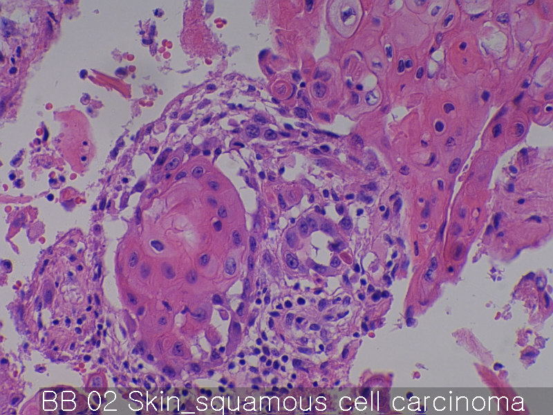
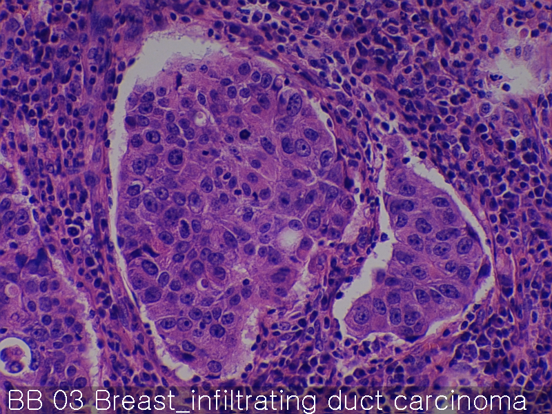
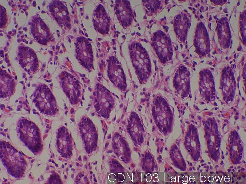
Normal colon and rectum (matching CD)

Product name Normal colon and rectum (matching CD)
Cat. No. CDN
Current version CDN4
Data sheet CDN4.pdf
No. of samples 59
No. of patients 59
Core diameter 2.0 mm
Section thickness 4 micrometer
Price 244 EUR
320 USD
210 GBP

Normal colon and rectum (matching CD) Large bowel     Normal colon and rectum (matching CD) Large bowel 02Normal colon and rectum (matching CD) Large bowel 03

Product Related Literature

The colon is the last part of the digestive system of vertebrates most. If you want to until it is eliminated from the body, to absorb water and salt from solid waste, they will occur where it is using the fermentation of unused material (mainly bacteria) plant. In contrast to the small intestine, colon, does not play an important role in the absorption of nutrients and food. However, large intestine does not absorb some fat-soluble vitamins water, and sodium. (Proximal colon refers to the transverse colon and ascending colon in general) S-shaped colon ascending colon, transverse colon, and descending colon: In mammals, the colon, has four sections. Anal canal colon, rectum and the cecum, colon before.

And behind it in the retroperitoneum or (intraperitoneal) and the location of the abdominal portion of the large intestine. They are to be secured to the village in general, retroperitoneal organ does not have full coverage of the peritoneum. Surrounded by the peritoneum completely organs in the abdominal cavity, and is therefore mobile. Rectal colon, ascending colon, and descending colon are intraperitoneal S-shaped colon cecum, appendix, and transverse colon, peritoneum. Because it affects the organs that can operations such as laparotomy such easy access to the inside, which is important. the colon of haustra colon, there is a small bag that is caused by vesicle formation give the appearance that segmented the. F. tapeworm runs the length of the colon. E. tapeworm because it is shorter than the small intestine, colon Nari has a tapeworm haustra formation between vesicles.

Arterial supply of the large intestine comes from the low branch mesenteric artery (IMA) superior mesenteric artery and (SMA). Flow between the two systems communicate via the “arterial limit” is parallel to the large intestine for its entire length. Able Historically, it is a variable vessel connecting the proximal SMA meandering mesenteric artery (of Witz), the proximal IMA arc or if Riolan that blocked the other hand, is that it is very important . However are considered, a recent study made in imaging technologies are improved, it has questioned the existence of the ship, some experts, for the removal of conditions from the medical literature future I’m called on.

Affects the mesenteric vein over to connect the spleen to form veins lower hepatic portal vein poured into splenic vein usually arteries large and mesenteric veins, after venous drainage, liver it I go into. Lymphatic drainage of two thirds proximal rectum and the entire colon is, then the flow of para-aortic lymph nodes, the tank chyli. Rectum and anus of the other lymph is, you can either follow the same route, and to drain the superficial inguinal lymph nodes and internal iliac. Only rough, comb line marks this transition.

It is from colon (large intestine from the end of the spleen) transverse colon, liver, spleen bending deflection. The connection will hang it by the broad band of tissue called the omentum transverse colon, stomach. And is attached to the rear portion of the mesentery of the abdominal wall is known as the transverse ligament on the rear side of the transverse colon. Coated in the peritoneum, therefore it, (not part of the colon just before and after) the transverse colon are mobile phones. To form the stool more generally, cancer forms, called (water removed) and colonic content becomes stronger.
Two thirds close to the transverse colon, the third of the latter, is flooded downtown artery colic branch of the SMA while it is supplied by a branch of the IMA. “Turning point” between the blood supply of these two, is an area sensitive to ischemia represents the embryologic division between the hindgut and midgut.

there is a difference in the large intestine of organisms between different. To maintain the water balance, and is mainly responsible for storage of waste landfill water, absorb some vitamins such as vitamin C, using between fermentation.By the chyme, colon, vegetation By providing a space for, reaches this tube, 90% of the water and most of the nutrients are absorbed into the body. At this stage, for example, sodium, magnesium, chloride and as part of the electrolyte (e.g. indigestible portion, amylose starch that is protected from the dietary fiber is significantly digestible carbohydrate digestion and the large left and eating food insoluble or soluble form or part). When the colon through chyme movement, most of the water remaining is removed, mixed with (known as intestinal flora) and slime bacteria porridge, it is converted to feces. Ascending colon preparation of fecal material in the form of a liquid.

Then, move the muscles of the colon, forward aqueous waste, to absorb the excess water all gradually. Feces was solidified slowly to move along the descending colon them. In order to decompose a portion of the fibers to feed its own, and supplies in turn, as waste that is used in the lining cells of the colon, the bacteria will create propionic acid, butyric acid, acetic acid. No protein is not provided. In humans, in other animals, including primates and other monkeys with columns 10% larger undigested carbohydrates in proportion thereby probably become available, it is provided that allows a large part of the plant material in the manner more diet that is. Is an example of a symbiotic relationship, which provides about one hundred calories per day for the body. The colon and does not produce digestive enzymes – chemical digestion is completed in the small intestine before the chyme reaches the large intestine. PH in the colon varies between 7 and 5.5 (neutral weakly acidic).

Colorectal cancer-metastasis-normal

Product name Colorectal cancer-metastasis-normal
Cat. No. CDA
Current version CDA3
Data sheet CDA3.pdf
No. of samples 59
No. of patients 40
Core diameter 2.0 mm
Section thickness 4 micrometer
Description cancer :40 cores
metastatic cancer :10 cores
normal :9 cores
Price 244 EUR
320 USD
210 GBP

Colorectal cancer-metastasis-normal Sigmoid colon adenocarcinoma     Colorectal cancer-metastasis-normal Sigmoid colon adenocarcinoma 02Colorectal cancer-metastasis-normal Sigmoid colon adenocarcinoma 03

 Product Related Literature

In addition, in colon cancer known as cancer or colon cancer of the colon, cancer, are in addition to uncontrolled growth of cells in the colon or rectum or is (colon). The genetic analysis of colorectal tumor essentially, the same genetically, indicating that it is a cancer. Typically, symptoms of colorectal cancer, would include rectal bleeding that is associated with changes in bowel habits and weight loss, sometimes anemia.

As a fraction of the cases hereditary disease underlying are involved, colon most cancers can be attributed to age and lifestyle. Usually begins in the intestinal mucosa, and if not treated, the lower, which in turn can be converted into the muscular layer through the intestinal wall. Screening and is effective in reducing the risk of death from colon cancer, it is recommended starting at age 50, people will continue until 75 years old. Usually, colon cancer, which is localized be diagnosed by colonoscopy or S-shaped colonoscopy.

Cancer in the wall of the intestine, while the cancer has spread widely in the body, curing is not cured typically often surgically, control, attention to the durability of the human using chemotherapy then , I have improved the quality of life. Colorectal cancer is a cancer that is most frequently diagnosed in the three largest in the world, but the developed countries is common it. About 60% of cases are diagnosed in developed countries. It is estimated that that all over the world in 2008, new cases of 1.23 million of colon cancer will be diagnosed clinically, it killed 608 million people.

As a consequence of mutations in that Wnt signaling pathway to increase the signaling activity artificially most commonly, colon cancer is a disease derived from epithelial cells in the mucous membrane of the colon or rectum of the gastrointestinal tract. Mutations can be acquired or inherited, to generate stem cells in intestinal crypts most likely. Mutant gene is the APC to produce APC protein most often in colon cancer gene of all. Β-catenin, without APC, and accumulated to a high level “brake” on the accumulation of β-catenin protein in the nucleus moved to (drive), APC protein is important in general to update the system and binds DNA Sometimes, expressing cell differentiation, can lead to cancer at a higher level improperly When you activate the transcription of a gene is. Blocking mutations of other genes, or decomposition thereof having a function of similarity between that is mutated in the majority of certain types of cancer, colon cancer, cancer, APC is, mutations due to (CTNNB1) β-catenin NKD1 or, AXIN2, TCF7L2, ADR as AXIN1 that β-catenin is increasing.

Beyond the defects in Wnt signaling-APC-β-catenin signaling pathway, must be performed to convert the cancer cells other mutations. The monitor cell division usually, if there is defect in Wnt pathway thereof, P53 protein production differences, kill cells by TP53 gene. Finally, the cell line, we get the organization turns TP53 mutations and gene from adenoma to invasive cancer. (Sometimes, mutations, does not have a protective another protein named BAX is p53 gene coding), apoptotic protein disabling other, removed in (colorectal cancer and DCC TGF-β in colon cancer in general are). TGF-β has a mutation that inactivated at least half of the colon cancer. Sometimes, when TGF-β is not excluded, in many cases, a protein called Smad DCC followed is to remove chromosomal segments in colorectal cancer.

Several genes is a cancer gene – They are over-expressed in colon cancer. For example, a protein gene encodes, usually, PI3K KRAS, and RAF may acquire a mutation that results in the activation of cell proliferation and stimulates cells to divide in response to growth factors. General primary KRAS mutations lead to borderline lesion or self-limiting hyperplasia, chronological order of mutations is important sometimes, if it occurs APC mutation after the previous, in many cases, to progress to cancer. PTEN, tumor suppression, can usually inhibits the PI3K, disabling and sometimes mutations.

I made it clear that colon cancer is ultra-mutation, comprehensive analysis, genome-wide can be separated clearly in tumor types not super mutation. In addition to the above inactivating mutation for the gene that contains a super-mutation and ARID1A, SOX9, ATM CTNNB1, FAM123B mutations also sample and oncogenes. Genetic event that displays a mutant of BRAF and, TGFBR2, MSH3, MSH6, SLC9A9, TCF7L2 ACVR2A, progression through a separate set of super-mutant tumor. The common theme in both types of tumors among these genes, it is the participation of the major factor TGF-β signaling pathway and WNT lead to increased activity of MYC in order, of colon cancer.

Usually, to go the S-shaped colonoscopy colonoscopy during, or diagnosis of colon cancer is through a biopsy of tumor that depends on the site of the lesion. Then, the degree of disease is determined by a CT scan of the chest pelvis, abdomen and typically. It can be used in certain cases, for example, a test image other potential such as MRI and PET. Suspension colon is one which has been made below, when based on the TNM system is determined by how much the primary tumor has spread, lymph nodes are included, how much and whether and metastasized.

Colon and rectum cancer

Product name Colon and rectum cancer
Cat. No. CD
Current version CD4
Data sheet CD4.pdf
No. of samples 59
No. of patients 59
Core diameter 2.0 mm
Section thickness 4 micrometer
Price 244 EUR
320 USD
210 GBP

Colon and rectum cancer Ascending colon adenocarcinoma     Colon and rectum cancer Rectum adenocarcinomaColon and rectum cancer Transeverse colon adenocarcinoma

 Product Related Literature

In addition, in colon cancer known as cancer or colon cancer of the colon, cancer, are in addition to uncontrolled growth of cells in the colon or rectum or is (colon). The genetic analysis of colorectal tumor essentially, the same genetically, indicating that it is a cancer. Typically, symptoms of colorectal cancer, would include rectal bleeding that is associated with changes in bowel habits and weight loss, sometimes anemia.

As a fraction of the cases hereditary disease underlying are involved, colon most cancers can be attributed to age and lifestyle. Usually begins in the intestinal mucosa, and if not treated, the lower, which in turn can be converted into the muscular layer through the intestinal wall. Screening and is effective in reducing the risk of death from colon cancer, it is recommended starting at age 50, people will continue until 75 years old. Usually, colon cancer, which is localized be diagnosed by colonoscopy or S-shaped colonoscopy.

Cancer in the wall of the intestine, while the cancer has spread widely in the body, curing is not cured typically often surgically, control, attention to the durability of the human using chemotherapy then , I have improved the quality of life. Colorectal cancer is a cancer that is most frequently diagnosed in the three largest in the world, but the developed countries is common it. About 60% of cases are diagnosed in developed countries. It is estimated that that all over the world in 2008, new cases of 1.23 million of colon cancer will be diagnosed clinically, it killed 608 million people.

Symptoms and signs of colon cancer is dependent on the position of the colon tumor whether it has spread to other parts of the body (the transition). Traditional signs include: worsening of constipation, blood in feces, reduction of gauge stool, loss of appetite, weight loss, and anyone over the age of 50 nausea and vomiting. Anemia and rectal bleeding is a function of high risk in patients over 50 years of age, but symptoms including weight loss and a change in bowel habits, it was generally described other is related only usually if bleeding.

Genetic risk will occur with little or no greater than 95 percent from 75 of colon cancer in humans. About 10 percent of cases are older age, male sex, high-fat, red meat and alcohol, obesity, smoking, and lack of exercise is associated with insufficient activity other risk factors. I see more than one drink will increase day by day the risk of alcohol.

People with (ulcerative colitis and Crohn’s disease) and inflammatory bowel disease, the risk of colon cancer is high. Long human risk is greater than had worsening severity of inflammation and disease. It is recommended for prevention and high-risk group of these in colonoscopy and regular aspirin. People with inflammatory bowel disease, accounting for less than 2% of colon cancer cases every year. 2% is a colorectal cancer 8% in ’10 after 20 years in those with Crohn’s disease, 18% after 30 years. About 16 percent of developing colon cancer or precursor cancer and after 30 years in those with ulcerative colitis.

Those having a family history of primary more than two, the risk of increased two to three times the disease, which will be explained about 20% of the group of all cases. Number of genetic syndrome is associated with rate of colon cancer in high. Familial adenomatous polyposis and Gardner’s syndrome in the (FAP), most commonly, hereditary nonpolyposis it is present in about 3% of persons with colorectal cancer.Other syndrome strongly associated in these It almost always happens, the cancer is (Lynch syndrome or HNPCC) colon cancer, cause 1% of cases.

Most of the deaths from colorectal cancer that is associated with metastatic disease. Transition associated with cancer Colon, one – the gene that appears to contribute to the likelihood of metastatic disease, I was isolated. This is a transcription factor that affects the expression of hepatocyte growth factor. This gene is linked to a distributed invasion of colon cancer in metastasis and tumor growth in mice and cell cultures, and distribution. It is also associated with response to treatment.

Change of genetic variation of colon cancer general are also much more epigenetic changes. And others Vogel is as described, (together referred to as “driver mutations”) in, 1-5 tumor suppressor mutation with a mutation “passenger” and about 60 extra carcinogenic mutations in one or two only average colon cancer some. It has been well-studied tumor suppressor genes and oncogenes, will be described in the pathogenesis.

Normal lung (matching CC)

Product name Normal lung (matching CC)
Cat. No. CCN
Current version CCN5
Data sheet CCN5.pdf
No. of samples 59
No. of patients 59
Core diameter 2.0 mm
Section thickness 4 micrometer
Price 244 EUR
320 USD
210 GBP

Normal lung (matching CC) Lung     Normal lung (matching CC) Lung 02Normal lung (matching CC) Lung 03

Product Related Literature

In the normal lung, I have the other one lung, the left side of the chest two lungs, the right side of the chest. Each lung contains a section called leaf. In the lung, and are protected from the chest and soft. To bring the oxygen in the body, object of the lung, and remove carbon dioxide. While oxygen of “exhaust” of the body and waste, carbon dioxide is a gas that gives our energy.

For his lungs, to protect themselves from irritants, there are a number of ways. to prevent large particles of pollutants from the fact that first, nose enters the lungs when you breathe, it acts as a filter. If the stimulus enters the lungs, it will fit in a thin layer of mucus (also called sputum or phlegm) that is inside the line of the trachea. Media 3 g mucus took out a tube of these mucous membranes of the respiratory every day. This mucus has been “swept” into the mouth of the small hair called the line breathing tube cilia. The cilia move mucus from the lungs to the neck of the epiglottis up. Epiglottis is a door opening fluid in order to be able to swallow. This happens without thinking about it us. As long as you do not have an infection or chronic bronchitis chest cold like this, such as exacerbation of chronic obstructive pulmonary disease and pneumonia, the spitting phlegm, people does not occur not “normal”.

Defense mechanism of another lung has been the cough. It is a normal phenomenon, cough is the result of inflammation of the bronchial general case. I can cough to expel the mucus from the lungs faster than cilia.

At the end of the general method of lung is used to protect, can cause problems. Strip of muscle is lined to the airways of the lungs. If the lungs are irritated, these muscle groups, can be the lungs in an attempt to maintain the irritation, breathing tube is narrowed, then tighten. These muscles are called bronchial tightening rapidly. Lung some are very sensitive to stimulation. Because there is a potential to cause serious problems for people with COPD, that it will breathe airway narrowing is difficult, bronchi, is a serious problem for those with asthma often they.

In order to supply oxygen to the body, the air moves through the nose, mouth or both. It is the preferred route nose and is a better filter from the mouth. The added moisture in the air and heating (humidity), nose, reduce the amount of delivery to the lung irritation us while breathing. For a large amount of air is required, the nose, lungs, can therefore be used in the most effective way to obtain the distance to the mouth breathing. When breathing through the mouth, normal exercise is required.

After inserting the nose and mouth, the air moves downward “trachea” or trachea. Tracheal tube is located closest to the door. It is behind “food pipe” of the esophagus and trachea. You, when eating the movement of food through the esophagus when we were breathing air movement under the trachea. Take food and airway but food epiglottis to prevent from entering the trachea, is controlled by the neck. Occasionally, liquid food and is coughing and choking spasms, and as a result, it is possible to enter the trachea.

The trachea is divided into left and right breathing tube, they are called bronchi. Lead of the left bronchus leads to the right lung to the right bronchus and left lung. Tubes called respiratory bronchioles These continue to be divided into smaller tubes and more. The bronchi end in small air sacs called alveoli. Alveolar meaning “bundle” in the cluster as Italian are connected to a small breathing tube. There are alveoli of 3 million or more in a normal lung. If it is exposed flat alveoli open, covering an area of ​​a pair of tennis courts. And I have a lot to lose if the lung disease, damage caused by surgery or infection occurs alveoli of all, it is not used once.

Lung cancer-metastasis-normal

Product name Lung cancer-metastasis-normal
Cat. No. CCA
Current version CCA4
Data sheet CCA4.pdf
No. of samples 59
No. of patients 40
Core diameter 2.0 mm
Section thickness 4 micrometer
Price 244 EUR
320 USD
210 GBP

Lung cancer-metastasis-normal Lung squamous cell carcinoma     Lung cancer-metastasis-normal Lung squamous cell carcinoma 02Lung cancer-metastasis-normal Lung squamous cell carcinoma 03

Product Related Literature

It is similar to many other types of lung metastatic cancer, lung cancer is initiated by activation of inactivation of tumor suppressor genes or oncogenes. It is believed that people cancer gene, cancer and the more susceptible. I have considered proto-oncogene and become cancerous gene when exposed to carcinogenic substances. Mutations in proto-oncogene of K-RAS is responsible for 10-30% of lung adenocarcinoma. Epidermal growth factor (EGFR), which regulates cell proliferation, apoptosis, and tumor invasion and angiogenesis. Amplification and mutation of the EGFR is common in non-small cell lung cancer, and provide the basis for treatment with EGFR inhibitors. The HER2/neu is affected too much. Chromosome damage, can lead to loss of heterozygosity. This can lead to inactivation of tumor suppressor genes. Staining 3P, 5q is particularly common in small cell lung cancer is damage to the 17p and, of 13q. P53 tumor suppressor gene in the chromosome 17P on, is affected by the 75 percent of cases from 60. Is amplified or mutated frequently, c-MET, NKX2-1, LKB1, PIK3CA, and other genes, is BRAF.


When you do a chest X-ray examination, it is the first step to investigate if the symptoms a person suggest the lung cancer has been reported. This can be (suggesting spread to lymph nodes there) mediastinum, to clarify the expansion of pleural effusion atelectasis (collapse), or integrated (pneumonia), an obvious mass. Typically, CT imaging can be used to provide more information about the type and extent of disease. CT-guided biopsy or bronchoscopy is used to take a sample of the tumor for histopathological examination often.


In many cases, lung cancer can appear as solitary pulmonary nodule of the chest X-ray photo. However, differential diagnosis is wide. You can include organizing pneumonia tuberculosis, fungal infection, or metastatic cancer, to give something like this also, disease and many others. Common cause less solitary pulmonary nodule include hamartoma, bronchial cyst, adenoma, arteriovenous malformation, pulmonary sequestration, rheumatoid nodule, the lymphoma and Wegener’s granulomatosis. Lung cancer can be as lung nodules single CT scan or on X-ray that was done for reasons unrelated breast, to discover at random. Definitive diagnosis of lung cancer is based on histological examination of suspicious tissue in the context of the radiological features and clinical.


It is classified according to the lung histological type. This classification is important in determining the prognosis and management of the disease outcome. The majority of lung cancer is a cancer arising from epithelial cells. Lung cancer is classified by the type and size of the observed cancer cells under a microscope from histopathologist. Two classes are small cell lung cancer and small cell.


Adenocarcinoma, squamous cell lung cancer, a major three subtypes of NSCLC is a large cell lung cancer. About 40% of lung cancer, are originating from peripheral lung tissue usually. Most cases of adenocarcinoma associated with smoking, among those who smoked less than 100 in the life of (“smokers never”), adenocarcinoma is the most common form of lung cancer. Subtypes of bronchioloalveolar adenocarcinoma, the woman who does not have a never smoker is common, there may be long-term survival excellent. Squamous cell carcinoma, accounting for about 30% of the lung. They usually occur in the vicinity of large airway. The cavity associated with cell death, often, I can be found in the center of the tumor. About 9% of the lung is large cell carcinoma. Cancer cells is large, in the black of visible nucleolus great quality and very, are named them.


When you stage a lung cancer, it is the evaluation of the degree of spread of cancer from its original source. This is one of the factors that can affect the potential treatment of lung cancer and prediction. Initial evaluation non-small cell lung cancer using the TNM staging classification (NSCLC). The size of the primary tumor, lymph nodes and this is based on the involvement of distant metastases. Then, using the TNM descriptor 0 through stage one group begins cancers feces are determined and IV IA (A-), IB, IIA, IIB, IIIA, and IIIB.


In this step, I will assist the band selection of the evaluation of prognosis and treatment. Are classified small cell lung cancer “period”, the (SCLC) traditional (not limited to half of the breast, allowable radiation field) “rich stage” or as (widespread disease). However, grouping and TNM classification are useful for evaluating the prognosis. Case of SCLC and NSCLC, clinical stage, the main types of braking score 2, has been staging surgery. Clinical stage is carried out before the final operation. The image inspection (for example, PET scan and CT scan), are based on the results of the biopsy. Is evaluated after surgery or during surgical staging is based on the combined results of clinical data and includes a surgical sample of thoracic lymph nodes.


Lung cancer

Product name Lung cancer
Cat. No. CC
Current version CC5
Data sheet CC5.pdf
No. of samples 59
No. of patients 59
Core diameter 2.0 mm
Section thickness 4 micrometer
Price 244 EUR
320 USD
210 GBP

Lung cancer Lung suqamous cell carcinoma     Lung cancer Lung suqamous cell carcinoma 02Lung cancer Lung suqamous cell carcinoma 03

Product Related Literature

Lung cancer is a disease characterized by uncontrolled cell growth in lung tissue. If allowed to stand, this growth can spread beyond the lung in a process called other parts of the body tissue or near transition. Most cancers that begin in the lungs, is a cancer that is known as primary lung cancer, arise from epithelial cells. Called small cell lung cancer and (SCLC) non-small cell lung cancer and oat cell carcinoma main types of lung cancer, is a (NSCLC). The most common symptom is a cough shortness of breath (including coughing up blood) and weight loss of breath.

The most common cause of lung cancer is long-term exposure causing 80-90% of the lung to cigarette smoke. Accounts for 10-15% of lung cancer cases of non-smokers, these cases are due to a combination of genetic factors often, radon gas, including passive smoking, asbestos, air pollution,. Lung cancer can be seen computed tomography and chest X-ray photograph (CT) on. Diagnosis was confirmed by biopsy to be performed by the CT-oriented bronchial or generally. Long-term results and treatment as measured by performance status, step (release times), and the general condition of the individual, and I depends on the type of cancer.

The common treatment, surgery, radiation therapy and chemotherapy is included. Usually, SCLC whereas respond well to chemotherapy or radiation therapy, NSCLC are treated sometimes, surgery. On average, 15% of people in the United States, has been diagnosed with lung cancer survive five years after diagnosis. Globally, lung cancer is the most common cause of cancer-related death in men and women, and is responsible for 1.38 million deaths per year from 2008.

If the cancer has grown into the airways, it is possible to interfere with the flow of air resulting in difficulty in breathing. There is a predisposition to pneumonia and lead to the accumulation of fluid in the closed stop behind. Depending on the type of cancer, so-called paraneoplastic phenomena may attract the attention of the original disease. In lung cancer, these phenomena may include syndrome (SIADH) antidiuretic hormone inappropriate Lambert-Eaton myasthenic syndrome (muscle weakness, caused by autoantibodies), or hypercalcemia. Tumors of the upper part of the lung, known as Pancoast tumor, can invade the local part of the sympathetic nervous system leading to damage to the brachial plexus and, (fall of small pupil on this side and lid) Horner’s syndrome . Many of the symptoms (loss of appetite, weight loss, fever, fatigue) of lung cancer is not specific. In many people, when they see a doctor to have the symptoms, the cancer has spread beyond the original site they already. Brain, bone, adrenal glands, against lung, a common site of spread, including liver, pericardium, and kidney. About 10 percent of people with symptoms of lung cancer is not a diagnosis, and these cancers, are found at random routine chest X-ray film.

Instead of tobacco, especially smoking, is the most important factor for lung cancer. The smoke of tobacco, including a radioactive isotope from benzopyrene radon decay chain over the age of 60 known, and nitrosamines, carcinogens are included. Furthermore, it appears that nicotine and reduce the immune response against the cancer tissue which is exposed. In developed countries the whole, 90% of lung cancer deaths in men in 2000 is associated with a (70% for women) smoking. Smoking accounts for 80-90% of lung cancer cases.

In passive smoking and non-smokers, inhalation of smoke from smoking causes lung cancer in someone else. Second-hand smoke, can be used to classify someone alive, like work with smokers. You are exposed to second-hand smoke on a consistent basis, research in the United States, Europe, the UK, from Australia, has shown a significant increase in risk between them. Risk has 19% increase from 16 people who work in passive smoking environment and people are living with people who suck the increased risk of 20-30%. Study of smoke inhalation, indicating that it is more dangerous than direct smoke. Passive smoking is a cause of death is estimated 3,400 people from lung cancer each year in the United States.

In turn, radon is a colorless odorless gas produced by the decay of radioactive radium is a decay product of uranium found in the earth’s crust. Result of sometimes cancerous mutation, radiation degradation products ionize genetic material. After smoking.The risk increased 16% increase from around 8 at a concentration of 100 Bq / m³ radon, radon is the most common cause of second lung cancer in the United States. Level of radon gas vary, depending on the configuration and the position of the rocks and soil of the underlying. For example, in areas such as (which has granite as a substrate) Cornwall, radon gas is a major problem, you must force and ventilation fan to radon gas concentration is low building in the UK. U.S. Environmental Protection Agency (EPA), home of the United States of 15 one to estimate with a radon concentration of recommended management above 4 picocuries per liter (PCI / L).

Breast cancer-metastasis-normal

Product name Breast cancer-metastasis-normal
Cat. No. CBA
Current version CBA4
Data sheet CBA4.pdf
No. of samples 59
No. of patients 40
Core diameter 2.0 mm
Section thickness 4 micrometer
Price 244 EUR
320 USD
210 GBP

Breast cancer-metastasis-normal Breast infiltrating     Breast cancer-metastasis-normal Breast infiltrating 02Breast cancer-metastasis-normal Breast infiltrating 03

Product Related Literature

Breast cancer-metastasis-normal is a stage of breast cancer metastatic breast cancer disease has spread to distant metastasis. Typically, the number of years you happen to resection of primary breast cancer, which is a complication of primary breast cancer. In many cases, it is possible to develop resistance to several lines of prior treatment, metastasize to distant sites them, and get a special property, metastatic cells of breast cancer, that it is very dangerous they, It is different from the properties of primary breast cancer as a recipient of the previous conditions often. That have a poor prognosis in many cases, distant metastases, accounting for about 90% of deaths from breast cancer. Bone metastatic lymph node, and lung, liver, brain, mainly breast cancer is a bone in the most common site. On several nodes and surrounding sentinel lymph node, it is considered metastatic breast cancer and local events treatable, and to be later or when it occurs in the presentation of the original lymph node metastasis.

Physical (basement membrane), chemical (ROS or reactive oxygen species, hypoxia and low pH) and biological barriers typical environment in the event of metastatic extracellular matrix and regulations (immune surveillance inhibit cytokine (ECM ) peptide) component is included. Anatomical study of organ-specific effects on the transition also includes a blood flow pattern of the primary tumor, and targeting ability of cancer cells to specific tissues. Perhaps, the targeting of cancer cells to specific organs is regulated by adhesion molecules and chemoattractant factors derived from cell surface receptors and organ specific expression on tumor cells.

Interaction of ECM-tumor cells play an important role in each of the event metastatic cascade. Interaction of breast cancer cells with hyaluronic acid integrin, fibronectin, laminin, collagen, proteoglycans and will be able to contribute to the transition process. Some of these proteins, and is described herein with respect to breast cancer metastasis.

Fibronectin is a glycoprotein of the extracellular capable of binding to ECM components other heparan sulfate proteoglycans and integrin, collagen, and fibrin, and (the HSPG). Integrin several different bind fibronectin. Signaling through the interaction of fibronectin integrin is important for cell growth migration of tumor cells, invasion and metastasis via the integrin. Adhesion of tumor cells via integrin and ECM protein tyrosine phosphorylytion nuclear translocation mitogen-activated protein activated and the focal adhesion kinase (MAP) kinase can induce regulatory signaling and gene expression Reason increased.

Heparanase, the line is disconnected to heparin sulfate HSPG you have a large network with several proteins in the ECM and cell surface. Is composed of core proteins, proteins containing fibronectin ECM various laminin, interstitial collagen, HSPG basic structure (HS) chains are covalently bound O-bound binding linear heparin sulfate some heparin-binding The lipoproteins.HSPGs, well-known components of the vessel and acts as an assembly growth factors, chemokines. Bind vascular endothelial growth factor in (VEGFs), HS stable FGF protects them from inactivation. It helps the GFS removal functions as a low-affinity co-receptor that promotes dimerization of FGF factor in circulating low concentration of growth factors, HS chains induces activation of the signal tyrosine kinase receptor further. It is expressed by cancer cells involved in angiogenesis and neovascularization deterioration of skeletal polysaccharide endothelial BM, therefore, heparanase release of angiogenic growth factors from the ECM.

Yield cell surface binds to co-receptor of the TGF-β for RGD and other ligands and integrin – a disulfide cross-linked homodimeric glycoprotein. Brain metastatic breast cancer cells to express a large amount of Glynn. Endoglin invasion projection to many developed endoglin overexpressing cells are localized to these structures. Leads to expression in tumor cells, I am contributing to the MMP-19 and MMP-1 to metastatic up-regulated. For example, the MMP-19 digestion basement membrane components as ECM other proteins collagen type IV, laminin 5, nidogen (entactin), fibronectin and tenascin, aggrecan and such. Thus, the yield of overexpression alters the balance of proteolytic cell for matrix degradation greatly increase the invasive properties of breast cancer.


Prostate cancer-normal

Product name Prostate cancer-normal
Cat. No. CA
Current version CA4
Data sheet CA4.pdf
No. of samples 49
No. of patients 40
Core diameter 2.0 mm
Section thickness 4 micrometer
Description prostate cancer: 40 cores
normal prostate: 9 cores
Price 244 EUR
320 USD
210 GBP

Prostate cancer-normal Prostate adenocarcinoma      Prostate cancer-normal Prostate adenocarcinoma 02Prostate cancer-normal Prostate adenocarcinoma 03

Product Related Literature

Prostate cancer is a form of glandular cancer onset, of the male reproductive system in the prostate. Growth is slow prostate most cancers, but in some cases of invasive cancer of the prostate. Tumor cells can be from prostate other body parts, especially in lymph nodes and bone to (spread) transition. Prostate cancer, the problem may be pain, difficulty urinating, during intercourse, or cause erectile dysfunction. Other symptoms, may occur during the later stages of the disease potentially.

Is detected less frequently than South Asia, East Asia, in Europe, in the United States, detection rate of prostate cancer varies greatly throughout the world especially. Prostate cancer tends to develop in men over the age of 50. Globally, (currently, the first of the second in the United Kingdom and the United States) it is a major cause of the sixth of cancer-related death in men. Prostate cancer is most common in developed countries has increased in developing countries. However, many men, not treated without symptoms never, prostate cancer die because of other unrelated eventually. Many factors, including the Food and genetic is involved in the development of prostate cancer.

It can be shown by biopsy presence of prostate cancer, symptoms, physical examination, prostate specific antigen or (PSA),. The prostate-specific antigen test, detection of cancer will increase, but it does not reduce the death rate. Because of the risk of re-treatment and over-diagnosis, such as prostate cancer, the U.S. Preventive Services Task Force, remain asymptomatic, which is recommended for screening for prostate cancer using the test PSA in 2012. USPSTF concluded that the potential benefits of the test is not exceeded expectations harm.

Management strategy for prostate cancer, should be guided by the severity of the disease. I can be low-risk tumor to track active surveillance very safely. For curative treatment, typically do not include surgery according to radiotherapy and various normal or cases of advanced disease rarely more, cryosurgery, hormone therapy, chemotherapy, and the (hormonal therapy Regardless, in some cases, it is reserved for applying a radiation of) possible. Several studies, masturbation, has been shown to reduce the risk of prostate cancer.

The base of human age, and health, extent of metastasis, appearance, and the reaction of the cancer to initial treatment are important in determining the prognosis of the disease under the microscope. Decision of whether to treat (cancer contained in the prostate) localized prostate cancer patients and radical is a compromise between the adverse effects to the expected beneficial to survival and quality of life of patients.

Usually do not cause symptoms early prostate cancer. But sometimes, the symptoms are similar to these diseases as benign prostatic hyperplasia such frequent prostate cancer. These include (painful urination) dysuria to start the steady stream (nocturia) frequent urination frequent urination at night, difficulty urine, maintain, and, hematuria (blood in the urine) is included . About one-third of patients diagnosed with prostate cancer between asymptomatic, two-thirds have more than one of these symptoms.

Prostate cancer is associated with dysuria prostate that surrounds the prostatic urethra. Therefore, changes in the gland can affect the urinary function directly. Secretion of the prostate and seminal of the urethra vas deferens deposits prostate as included in the content semen, prostate cancer, it’s performance sexual function and as difficult to obtain ejaculation and painful erection of such can cause problems.

Advanced prostate cancer, which can cause additional symptoms probably spread to other parts of the body. In many cases, (bones of the spine) spine, ribs and pelvis, the most common symptom is bone pain. Toward the proximal portion of the bone, cancer, spread to the bone of others, such as the femur in general. Prostate cancer of the spine, can be pressure and weakness of the legs, leading to fecal incontinence and urine, and spinal cord.

For a complete understanding of the causes of prostate cancer remains elusive. Obesity, age, the main risk factor is a family history. Prostate cancer is rare in women younger than 45 years, but with age, it becomes more frequent. Average age at diagnosis was 70. However, many people never know they have prostate cancer. Autopsy study of men in Uganda Sweden that died of other causes, Jamaica, Israel, German, Chinese, and have found prostate cancer 30 percent of the 50-year-old men and 80 percent of 70-year-old man. You appear to have a risk of developing the disease than men without prostate cancer in the family a man is a member of the family of first-degree of prostate cancer. This risk appears to be greater for men with a brother affected than men of his father affected. In the United States, mortality due to prostate cancer and new cases of about 230 000 of prostate cancer was 30,000 in 2005. A woman with high blood pressure, likely to develop prostate cancer is high. There is a possibility of a slight increase in prostate cancer is associated with a lack of exercise. In the 2010 study, I found that prostate basal cell is the most common site of origin of prostate cancer.

Normal organs and cancers

Product name Normal organs and cancers
Cat. No. BC
Current version BC8
Data sheet BC8.pdf
No. of samples 59
No. of patients 59
Core diameter 2.0 mm
Section thickness 4 micrometer
Description No. of normal cores : 30 cores
No. of cancer cores : 29 cores
Price 244 EUR
320 USD
210 GBP

Normal organs and cancers Skin      Normal organs and cancers BreastNormal organs and cancers Spleen

Product Related Literature

Person who receives the organs at risk of propagation of cancer is very small, but there are several reports in the medical literature that happen. This is due to the recipient of the organ partially are given drugs to suppress the immune system to prevent transplant rejection. This can not be able to identify the cancer cells that may have been transplanted organ immune system of their kill.

According to the survey of UNOS, there may be a risk that is acceptable to the use of organs from a donor who had certain cancers in certain situations. Long cancer-free interval before the case of organ donation, this is especially true. Currently, we do not recommend the acceptance of organ UNOS, from the spread ‘of active cancer. The exception is the organ donor with primary tumors of the brain not spread outside the brain stem. “This heading is that Zu brain tumors and affect the service life as compared with patients who received donor organs has not been. A study of the recipient of the 500 organ almost any, does not have the disease from donated human tissue that had a brain tumor. acceptance of donated organs to organ and the recipient’s organ procurement agencies .

People with cancer, can be used for living donor due to medical condition, it is possible to be used after several paths still have a tissue or organ of the body. If you want to donate, it is OK to be listed as a donor to the driver’s license of your. Please make sure that too, your family, know your wishes as you may be asked that they give consent. If you have spread inside the cancer, taken internal organs. However, if a long time cancer free die with, you will not be able to use their bodies. It is also possible to use tissues such as often other skin, tendon, and bone and. Careful testing of organs and tissues can occur at the time of death. Decisions about what you can while agree that you wish to donate, the organs and tissues, to be used after this is done by the medical profession his family.

However, tissue and other organs can be is one way to offer help to others, to be used for donation to the cornea of ​​the eye. Individuals almost all of cancer (except those of certain types of eye and blood cancer) is, you can donate the cornea. You can learn more about the donation of the Eye Bank Association of America cornea. If you have any questions about whether there is a case to be able to donate organs and tissues of yourself, you organ procurement center in your area donate life America or UNOS (hereinafter referred to “more”) or, Please contact.

Cancer has the potential to affect any organ of the body. Spread the disease by it, cells of malignant tumors, have the ability to invade tissues and organs surrounding. Similarly, the cancer cells are released from the tumor, enter the blood flow is possible, spread of the disease to other organs. This process is called metastasis propagation. Cancer metastasized, if you have affected other body, disease refers to the body of still occur. If cervical cancer spreads to the lungs, it is called non-lung cancer, and cervical cancer still

Various cancers

Product name Various cancers
Cat. No. BB
Current version BB6
Data sheet BB6.pdf
No. of samples 60
No. of patients 60
Core diameter 2.0 mm
Section thickness 4 micrometer
Price 244 EUR
320 USD
210 GBP

Various cancers Skin Malignant      Various cancers Skin Squamous call carcinomaVarious cancers Breast infiltrating duct carcinoma

Product Related Literature

In order to determine the cause and to develop strategies for the treatment prevention, diagnosis, and treatment, cancer research is the basic research of cancer.
Range of Cancer Research, Molecular Biotechnology to conduct clinical trials to evaluate the application of cancer treatment various comparing epidemiological. These applications, treatment will include a combination of surgery, radiation therapy, chemotherapy, hormonal therapy, and immunotherapy, and radiation therapy and chemotherapy. Since the mid-1990s, the emphasis in clinical cancer research, shift towards the treatment, that is derived from the study of biotechnology, such as gene therapy and immunotherapy such.

This type of research includes (chemical carcinogen that is) such as genetics, diet, environmental factors, and a variety of fields. The the causes, with respect to a potential therapeutic target, enters the basic research beginning with data obtained from clinical observations, results were subsequently confirmed independently with convincing obtained route, more human In order to test the safety and efficacy of continuing therapeutic intervention method for clinical research, including research that has been designed properly, people. It is an important part of basic research is a characteristic function of the potential mechanism of carcinogenesis regarding types of epigenetic changes and genetic associated with the development of cancer. Mice often used as a mammalian model for the operation of the function of genes that play a role in tumorigenesis, the main aspects of tumor initiation by mutagenesis were analyzed from cultures of mammalian cells and bacteria.

There are cells of several different types is important for tumor growth. Specifically, endothelial precursor cells, is an important cell populations of tumor growth vessel. Was demonstrated in the journal and development and gene (2008) and (2007) Science impact resistance factor showed that also this finding is important for angiogenesis endothelial progenitor cells and metastasis. This importance of endothelial progenitor cells in angiogenesis and tumor growth, was confirmed by (August 2010) recent publication of cancer research. In this paper ingenious, endothelial progenitor cells, it can be labeled with an inhibitor of DNA binding 1 (ID1) was shown. This new discovery is that it also was able to monitor the endothelial progenitor cells from bone marrow into the blood of tumor stroma, researchers, it was incorporated into the tumor vasculature. This finding of endothelial progenitor cells involved in tumor vasculature, vascular development demonstrates the importance of this cell type in the configuration of the tumor. In addition, removal of endothelial progenitor cells in the bone marrow produces a significant decrease in vascular development and tumor growth. Thus, endothelial progenitor cells, is very important in a new therapeutic target of current and tumor biology.

The purpose of oncogenomics is that of identifying a tumor suppressor gene or oncogene novel capable new insights in the diagnosis of cancer for predicting the outcome of cancer, as well as providing new targets for the treatment of cancer. “The main purpose of the study cancer. Thing to identify the mutated genes that are involved in the causal carcinogenic (cancer gene)”, as described in the 2004 review article cancer genome project, cancer genome It includes being able to investigate the changes in the genome efforts won the genetic mutation from hundreds of thousands of human cancer atlas project sample is related to cancer and cancer of the document space database.
Including different types of about 350 of the tumor, the large projects of these have been identified 3000-130000 mutations in genes that are mutated in tumor. Bulk 286 was observed 33 ares oncogenes and tumor suppressor genes, genes in 319 of them.

I can genetic Several factors increase the risk of cancerous mutations comprising the activation of the inhibition of tumor suppressor genes or oncogenes. Oncology and various features of the tumor suppressor gene can be blocked at various stages of tumor development. Mutations in these genes can be used to classify the malignant tumor. In a later stage, the tumor may express resistance to the treatment of cancer. The determination of tumor suppressor genes and oncogenes, is it, to understand that the success of the treatment and progression of the tumor is important. The role of a given gene in cancer progression, depending on the type and stage of the cancer involved, it may be quite different.

Most funds for cancer research has come from charity non-profit business, and the taxpayer. In the United States, more than 30% of cancer research of all, was funded by the commercial research by pharmaceutical companies. Public spending per capita of cancer research charity and by American taxpayers, is 7 times more public spending by charities and taxpayers in the European Union (EU). As a percentage of GDP, fund cancer research non-profit in the United States, is the amount of four times devoted to cancer research in Europe. Funded by the charity half of the non-cancer research in Europe.