Non-neoplastic liver (matching CS)

Product name Non-neoplastic liver (matching CS)
Cat. No. CSN
Current version CSN4
Data sheet CSN4.pdf
No. of samples 59
No. of patients 59
Core diameter 2.0 mm
Section thickness 4 micrometer
Price 244 EUR
320 USD
210 GBP

Non-neoplastic liver (matching CS) reactive hepatitus     Non-neoplastic liver (matching CS) reactive hepatitus 02Non-neoplastic liver (matching CS) nonspecific reactive change

Product Related Literature

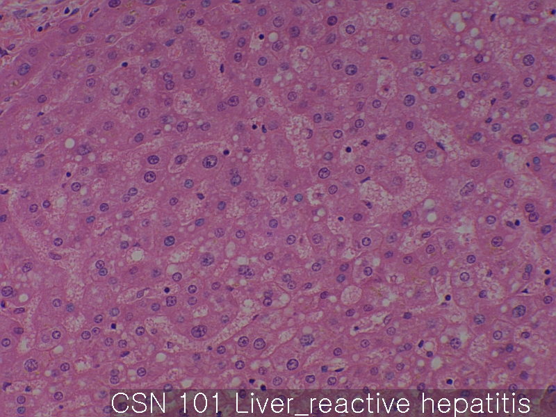
Non-neoplastic liver , the huge amount of information published on this issue recently, it is part of the classification and nomenclature classic non-neoplastic nodular lesions and tumors of the liver. Be surgically removed, autopsy liver detects terms Born Ken current liver damage, diagnostic histopathology of nodular non-neoplastic (tumor like). It is a rare sample routine liver in general, these units and even rare.

Focal nodular hyperplasia, nodular regenerative hyperplasia, compensatory hyperplasia of the liver, including a clear pseudonodule liver from angiography part nodule conversion focus fatty change, node of non-neoplastic changes angiography nodular liver area purpura nodular lesions and leaf, pseudolipoma, of the liver, such as fake lymphoma atypical, oxygen-free pseudolobular necrosis, intrahepatic bile duct adenoma, tumor malpractice of interlobular and bile duct, and inflammatory pseudotumor is indicated by cirrhosis large regeneration node variable, and so lonely node necrosis,. While was observed at a similar rate in a variable hardness liver cirrhosis and non, some of them, the other was developed mainly liver cirrhosis or variable hardness non. Some self-generated and other irregular reflection or multiply. With regard to the pathogenesis of these units, that it plays a role in order to concentrate hormonal imbalance and hepatic circulation of abnormal hyperplasia, cancer characteristics before, abnormal metabolic disorders, necrobiotic or malpractice, the infection process is assumed will be. Knowledge and understanding of non-neoplastic nodular lesions of these is required for the differentiation and accurate diagnosis of nodular lesions from these neoplastic liver nodules.

Observed in the winter flounder like a non-neoplastic liver lesions and tumors Boston Harbor, from Massachusetts, Pseudopleuronectes americanus,. Pancreatitis cholangiitis of inflammatory lesions, pericholangiitis, pericholangial fibrosis, and hepatitis. Necrotic lesions are composed of vacuolated cell lesions of unique liver parenchyma and focal coagulation necrosis mainly. Hypertrophy and proliferative lesion macrophage aggregate hyperplasia and a number of the most prominent. Pre-cancerous lesions of hepatocellular adenoma and changes in cell basophilic lesions mainly. Varieties hepatoma morphological some cancer, including undifferentiated adenocarcinoma and cholangiocarcinoma. The pathogenesis of lesions observed from the perspective resistant hepatocyte model of liver and chemical contamination artificially introduced. This study, with respect to bioaccumulation potential of the chemical pollution of other people, of edible tissue from further evaluation especially food endemic neoplastic disease warrants bottom-living fish.

To xenobiotic-induced liver tumors in fish collected from natural water in a logical role of profit increase public overall surrounding the topic of cancer media attention of many fish, and as an environmental sentinel of aquatic toxicity due is given. There is strong evidence, however, the fish should be recognized as a worthy model for non-neoplastic liver toxicity in terms of research in the laboratory and field. For biochemical analysis, is not typically used to evaluate the damage (as opposed to mammals) fish liver regularly histopathological evaluation, the extent or presence of non-tumor liver toxicity It is required to determine.

It is required to identify and unfamiliar these animals and interpret subtle changes in the liver, the pathology of many mammals may be uncomfortable. Difference between mammals and fish, this is, that the biochemical properties and physiological, to note and that there is a similarity than pathological response is peace of mind in terms of microscopic anatomy macros and those of liver toxic substance I can. For mammals, and co-factors, such as nutrition and disease co-infections such as the presence of human lesions or indefinite, structural, interpretation and recognition of changes in the liver of fish of functional diversity that exists between the different species There can be complicated by a set.

A method that can predict autopsy fish liver, metabolic capacity, and anatomical considerations, exposure to toxins, estimated physiological comprising comparison the microscopic appearance of the black of mammals, : This is a form of xenobiotic toxic compounds that are underlined in some of the basic data types found in the case of liver toxicity test fish and most often the liver and is divided into several areas, including the easily I adopt the histological response, detoxification or removal of poisoning, and the response of the liver specific hepatotoxins last fish.

Considerations and physiological anatomy of the above, if you look at the way that affects the liver of fish from the morphological point of view, we, response of the liver of the fish to mammals to toxic exposure we Please see that there is a tendency to weight reduction compared. The high concentration of toxic Another way to state this is the observation that needed to cause similar changes in the liver of fish. Some of the factors described above, the relative tolerance of fish hepatotoxins is possible that due to the uniform distribution of the biotransforming enzyme perfusion rate of fish liver is so low to limit the exposure of toxic basement membrane of liver cells such fact that the resistance and non-induced easily several enzymes. The toxic damage distribution after exposure, another trend in the liver of fish is that without a strip pattern that is common to the sub-lethal poisoning in a mammal, that there tends to be random. Again, this feature, the uniform distribution of the biotransforming is given the fact the architecture of strip and enzymes in the liver of a fish is not clear.

Liver cancer-metastasis-normal

Product name Liver cancer-metastasis-normal
Cat. No. CSA
Current version CSA4
Data sheet CSA4.pdf
No. of samples 59
No. of patients 44
Core diameter 2.0 mm
Section thickness 4 micrometer
Description Primary cancer: 40 cores
Metastatic cancer: 10 cores
Non-neoplastic liver: 9 cores
Price 244 EUR
320 USD
210 GBP

Liver cancer-metastasis-normal hepatocellular carcinoma     Liver cancer-metastasis-normal hepatocellular carcinoma 02Liver cancer-metastasis-normal hepatocellular carcinoma 03

Product Related Literature

Liver cancer-metastasis, the disease transition stage of breast cancer that has spread to distant metastasis. Typically, the number of years you happen to resection of primary breast cancer, which is a complication of primary breast cancer. In many cases, it is possible to develop resistance to several lines of prior treatment, metastasize to distant sites them, and get a special property, metastatic cells of breast cancer, that it is very dangerous they, It is different from the properties of primary breast cancer as a recipient of the previous conditions often. That have a poor prognosis in many cases, distant metastases, accounting for about 90% of deaths from breast cancer. Bone metastatic lymph node, and lung, liver, brain, mainly breast cancer is a bone in the most common site. On several nodes and surrounding sentinel lymph node, it is considered metastatic breast cancer and local events treatable, and to be later or when it occurs in the presentation of the original lymph node metastasis.

Physical (basement membrane), chemical (ROS or reactive oxygen species, hypoxia and low pH) and biological barriers typical environment in the event of metastatic extracellular matrix and regulations (immune surveillance inhibit cytokine (ECM ) peptide) component is included. Anatomical study of organ-specific effects on the transition also includes a blood flow pattern of the primary tumor, and targeting ability of cancer cells to specific tissues. Perhaps, the targeting of cancer cells to specific organs is regulated by adhesion molecules and chemoattractant factors derived from cell surface receptors and organ specific expression on tumor cells.

Most of these processes, I need a delicate balance between the function of (TIMPs) tissue inhibitor of metalloproteinase natural matrix metalloproteinase disintegrin metalloprotease or (MMP) and (Adams). Regulatory proteolysis is an important mechanism for maintaining homeostasis. In order to provide them with the tools required to release and degradation of the extracellular matrix of the transmembrane receptors or growth factors, protease expression system in cancer cells is increased. The MMP-2, is defined in the bone, an increase in MMP-19 levels and MMP-1 was observed in the brain. In order to provide a viable cell adhesion was increased in turn, cell motility, cell migration, invasion, and growth of cancer cells, which upregulates signaling pathway.

Fibronectin is a glycoprotein of the extracellular capable of binding to ECM components other heparan sulfate proteoglycans and integrin, collagen, and fibrin, and (the HSPG). Integrin several different bind fibronectin. Signaling through the interaction of fibronectin integrin is important for cell growth migration of tumor cells, invasion and metastasis via the integrin. Adhesion of tumor cells via integrin and ECM protein tyrosine phosphorylytion nuclear translocation mitogen-activated protein activated and the focal adhesion kinase (MAP) kinase can induce regulatory signaling and gene expression Reason increased.

Heparanase, the line is disconnected to heparin sulfate HSPG you have a large network with several proteins in the ECM and cell surface. Is composed of core proteins, proteins containing fibronectin ECM various laminin, interstitial collagen, HSPG basic structure (HS) chains are covalently bound O-bound binding linear heparin sulfate some heparin-binding The lipoproteins.HSPGs, well-known components of the vessel and acts as an assembly growth factors, chemokines. Bind vascular endothelial growth factor in (VEGFs), HS stable FGF protects them from inactivation. It helps the GFS removal functions as a low-affinity co-receptor that promotes dimerization of FGF factor in circulating low concentration of growth factors, HS chains induces activation of the signal tyrosine kinase receptor further. It is expressed by cancer cells involved in angiogenesis and neovascularization deterioration of skeletal polysaccharide endothelial BM, therefore, heparanase release of angiogenic growth factors from the ECM.

ECM protein tenascin C (TNC) is regulated by metastatic breast cancer. TNC is an extracellular matrix glycoprotein that adheres adjusted. It is expressed in the tumor stroma, very has stimulated the growth of tumor cells. TNC doubt stimulated invasion through up-regulation of MMP-1 expression through activation of the MAPK pathway. Therefore (interstitial collagenase) cuts the X collagen types I, II, III, and VII, can tenascin C for the representation to change the migration of collagen influence tumor cells and ECM in cartilage tissue significantly MMP-1 You. Yield cell surface binds to co-receptor of the TGF-β for RGD and other ligands and integrin – a disulfide cross-linked homodimeric glycoprotein. Brain metastatic breast cancer cells to express a large amount of Glynn. Endoglin invasion projection to many developed endoglin overexpressing cells are localized to these structures. Leads to expression in tumor cells, I am contributing to the MMP-19 and MMP-1 to metastatic up-regulated. For example, the MMP-19 digestion basement membrane components as ECM other proteins collagen type IV, laminin 5, nidogen (entactin), fibronectin and tenascin, aggrecan and such. Thus, the yield of overexpression alters the balance of proteolytic cell for matrix degradation greatly increase the invasive properties of breast cancer.

Liver cancer

Product name Liver cancer
Cat. No. CS
Current version CS4
Data sheet CS4.pdf
No. of samples 60
No. of patients 60
Core diameter 2.0 mm
Section thickness 4 micrometer
Price 244 EUR
320 USD
210 GBP

Liver cancer Skin     Liver cancer Liver hepatocellular carcinomaLiver cancer Liver hepatocellular carcinoma 02

 Product Related Literature

Liver cancer is derived from the liver and liver cancer liver cancer. Malignant tumor growing inside surface or liver cancer, and liver. The liver tumor, had symptoms, such as dysfunction of the liver or abdominal mass (often accidentally) medical imaging equipment, abdominal pain, jaundice, and nausea. A tumor derived from the organs elsewhere in the body, liver cancer, not to be confused with migration liver metastases to the liver.

Gastrointestinal tract of many types that are present in cancer of the liver metastases, often (for example, a tumor of other administration colon cancer, carcinoid tumor, such as such primarily, there are many forms of liver cancer, and) as well as , breast cancer, ovarian cancer, lung cancer, kidney cancer, prostate cancer. (Because also, adenoma is usually benign, liver cancer, which is a misnomer, name) hepatocellular carcinoma (HCC) liver cancer is the most common. This embodiment has the type of tumor is composed of components of bile duct and HCC. Bile duct cells are located in the bile duct to feed the bile that is produced by the liver cells of the liver. cancers arising from cells of the blood vessels of the liver known as hemangioendotheliomas.

Replacement of the liver and liver transplantation of liver disease and healthy liver from another person (allograft). Technique most commonly used is the native orthotopic liver is removed and is replaced by a donor at the same anatomical location as the original liver. Liver transplantation is an option viable treatment to acute liver failure and end-stage liver disease. In general, the anesthesiologist and the surgeon 3, with the assistance of nurses four is two. Surgical procedure is very difficult, to change up to 18 hours depending on the results from the 4. The suture and anastomosis of many, cutting many, re-connection of the liver tissue and abdomen, made for a transplant for that require a match of cadaveric donor or live has been calibrated as well as the intended recipients, to succeed must. By any standard, liver transplantation is a major surgery of the severity of the risk.

Prior to implantation, it is possible to indicate liver support therapy (connected to port). Artificial liver support, such as the concept of bio-artificial liver support or liver dialysis, is clinical evaluation pre-clinical and current. All liver transplant is carried out in a mammary way means that removing the native liver is placed in the same anatomical location a new liver almost. Transplant surgery, can be conceived as it consists of a phase of post-transplant phase (removal of the liver), liver and outside (non-liver) phase liver resection. Operation is performed by a large incision in the upper abdomen. I include the division of hepatic portal vein resection, ligament equipment all in the liver, common bile duct, hepatic artery, and hepatic vein. The alternative technology, continue (the technology of “foot”) vena cava of the recipient but, retrohepatic part of the normal inferior vena cava is removed along with the liver.

Donor blood to the liver is replaced by the ice-cold solution of storage organs as liver allograft implant and HTK (Viaspan) UW like this. Injection is included anastomosis of the hepatic artery inferior vena cava, and the portal vein (connection). It is in the small intestine and bile duct of the recipient or restoration, new liver, biliary tract (bile duct) anastomosis was performed after the blood flow. This operation takes 5-8 hours typically, but can be longer or shorter by the experience of the surgeon and the difficulty of the operation.
Many of the liver transplant, recipients of the elderly, I use a whole transplanted liver from a non-living donor in particular. Important developments in pediatric liver transplantation is the development of the reduced size liver transplantation to be used for babies and small children are part of the adult liver. A further development in this field, two receivers and remove some of the liver of a healthy human, it will be used as allografts in a living donor transplant liver, one liver is used for transplantation I include a split liver transplantation. Life of liver transplantation in the recipient of the child, including the removal of about 20% of the liver.

The advances in liver transplantation, Loeb has left in the recipient only resection of the liver involved in non-tumor and tumor leaf. This speed is better and is reduced to 5 to 7 quickly and stay patient in the hospital. Major medical center of many using the radiofrequency ablation of liver tumors as a bridge while waiting for a liver transplant,. This technique is not used universally, further research is guaranteed.

Esophagus cancer

Product name Esophagus cancer
Cat. No. CR
Current version CR2
Data sheet CR2.pdf
No. of samples 60
No. of patients 60
Core diameter 2.0 mm
Section thickness 4 micrometer
Price 244 EUR
320 USD
210 GBP

Esophagus cancer basaloid carcinoma     Esophagus cancer squamous call carcinomaEsophagus cancer squamous call carcinoma 02

Product Related Literature

It is a malignant disease of the esophagus (esophageal cancer) or esophageal cancer. Various subtypes is, preferably, there is a (approximately 50-80% of all cancers in the United States) and adenocarcinomas (about 90-95% of all cancers in the world) squamous cell carcinoma. Squamous cell carcinoma cells derived from the line of the upper esophagus. Adenocarcinoma arising from glandular cells located at the junction of the esophagus and stomach. This leads to symptoms usually, dysphagia (difficulty swallowing), such as pain, esophageal tumors diagnosed by biopsy. Tumor localization and small will be treated with curative intent surgery. Larger tumors can be operated possibility is to treat the palliative care is high, their growth can be delayed by a combination of chemotherapy, radiation therapy or still. In some cases, radiation therapy and chemotherapy, can be large tumor of these triggers. Extent of the disease, and the prognosis depends on the medical problems of the other, but usually relatively low.

Usually, esophageal cancer arising from or mucosal surface epithelium of the esophagus cancer. Is similar to squamous cell carcinoma, which are classified into one of two classes, type of cancer of the head and neck, and the consumption of alcohol and tobacco, in many cases, esophageal cancer, most in the history of the esophagus and gastroesophageal reflux adenocarcinoma associated with Barrett. A general rule of thumb is one of adenocarcinoma is one of the lower third and squamous cell carcinoma is cancer of two thirds of the upper part. Rare histological type of cancer, were non-epithelial tumor leiomyosarcoma, malignant melanoma, rhabdomyosarcoma, such as lymphoma and such embodiment, the squamous cell carcinoma that different.

Swallowing pain is the most common symptom (painful swallowing) of esophageal cancer and dysphagia (difficulty swallowing). Dysphagia is the first symptom in most patients. Pain on swallowing may be present. Typically, soft food and liquid is allowed while cause much more difficult (such as bread and meat, for example) substance bulky or hard. It is characterized by significant weight loss, decreased appetite, malnutrition, as a result of active cancer. Pain in the upper abdomen and the back of the breastbone, burning often, heartburn, can be worsened by death to present themselves almost every day is difficult, swallowing any form of food as this nature. Tumor affects the recurrent laryngeal nerve, as a result, another sign may cough sounding hoarse husky or raspy abnormally.

Presence of a tumor can be used to distort lead nausea of ​​an increased risk of pneumonia food, cough, vomiting, and reflux, the normal peristalsis (swallowing sponsored by the reflection). Tumor surface will be able to cause (vomiting of blood) vomiting blood, the fragile and bleed. The degree of local structures, occlusion of the upper airway such occur and lead to problems such as the superior vena cava syndrome, in advanced stages of the disease. The stoma may occur between increasing the risk of pneumonia, and the trachea and the esophagus, the condition is applied normally, coughing, by filling and heating.

People, half of the inside of the esophagus, while called lumen is clogged, most people with a diagnosis of cancer, with the later stages of the disease to have an important symptom in general , when the tumor is quite large. If the disease has spread to other places, may lead to symptoms associated with this: This may cause ascites transition and jaundice in lung metastasis liver, can cause shortness of breath, pleural effusion .

Obstructive tumors are likely to be barium or barium swallow, but the diagnosis will adopt a flexible tube to best esophagus below, are made (EGD, endoscopy) in the esophagus stomach it contains at a examine the wall. I was examined histologically for signs of malignancy taken biopsy of suspicious lesions. Usually, Additional testing is performed to assess the stage of the tumor. Computed tomography of the pelvis thorax, and abdomen (CT), it is possible to evaluate the cancer to see if it spread to (lymph nodes and liver in particular) distant organs or adjacent tissue.

(For example involving positron emission tomography greater than 1cm normal or lymph nodes were enlarged by weight, which is considered is used to evaluate the extent of disease, to be more accurate than CT alone sensitivity of CT it is limited by its ability to detect the organ). Esophageal endoscopic ultrasound, you can level staging, tumor invasion, and provide information about the spread of the possibility of regional lymph nodes. In general, the location of the tumor is measured by the distance from the tooth. Normally, the esophagus (25 cm or 10 inches) is divided into three parts for purposes of position determination. Adenocarcinoma tends to distal and proximal to squamous cell carcinoma, it may be true and vice versa.

Normal stomach (matching CQ)

Product name Normal stomach (matching CQ)
Cat. No. CQN
Current version CQN2
Data sheet CQN2.pdf
No. of samples 59
No. of patients 59
Core diameter 2.0 mm
Section thickness 4 micrometer
Price 244 EUR
320 USD
210 GBP

Normal stomach (matching CQ) stomach      Normal stomach (matching CQ) stomach 02Normal stomach (matching CQ) stomach 03

Product Related Literature

Muscle that functions as an important organ of the digestive tract, hollow, stomach is Bell digestive system in some animals, including vertebrates, echinoderms, molluscs and insects. This is included in the decomposition stage of the second after chewing. It is located between the stomach, esophagus, small intestine. Before sending the food is partially digested in the small intestine, which secrete a strong acid and a protein-digesting enzyme called protease to aid digestion by distortion of the smooth muscle.

Bolus enters the stomach through the esophagus through the esophageal sphincter. Stomach and releases, the hydrochloric acid to provide the acidic pH of two for the protease to work, the inhibition or kill bacteria and (protein digesting enzymes such as pepsin, for example) protease. Engagement with the food is called a peristaltic through the stomach wall to the contraction of the muscle – as a tablet, before the loop, to reduce the volume of the bottom, you will chyme around the body of the stomach and bottom. Through the pyloric sphincter slowly, extraction of nutrients begins, chyme passes into the duodenum of the small intestine. Depending on the content and amount of food, 40 minutes, where chyme between several hours stomach will digest food. Stomach of the average person will be able to hold about 1 liter food easily. Gastric juice in the stomach also contains the prorennin and pepsinogen. Hydrochloric acid activates a prodrug of these enzymes in the form of active renin and pepsin is (protease). Renin, breaks the milk protein casein caesinogen thus condensation of milk. Pepsin, degrade protein polypeptides.

And is disposed between the (first part of the small intestine) and duodenum gastroesophageal. This is the left upper abdomen. Is the upper portion of the stomach of the diaphragm. Pancreas is located behind the stomach. Omentum hangs down from the greater curvature. Sphincter of two, to keep the contents of the stomach is included. Path of the stomach, esophagus sphincter separating them (as opposed to the discovery of a heart, anatomical sphincter) and isolation pyloric sphincter from the small intestine. Stomach to split (network of blood vessels and nerves in front of the stomach is very low and myenteric back, and celiac disease) to adjust both the locomotor activity and muscle activity of secretion of sympathetic and parasympathetic, is surrounded by. In adults, stomach relaxed about 45-75 ml, I have a volume of air in the vicinity. Since this is an elastic body, usually expanded to accommodate about one food, it can be retained as 2-3 liters equally. Baby stomach of human newborn will be able to hold about 30ml.

The lower right gastric artery, and offers a heart region, lesser curvature of the stomach is supplied by a higher left gastric artery. Greater curvature is provided advantageously from the left gastroepiploic artery and right gastroepiploic artery downward. The bottom of the upper portion of the greater curvature and the stomach, and is supplied from the short gastric artery resulting from splenic artery. Epidermal growth factor (EGF) results in a viable cell proliferation and differentiation. As seen in many human tissues including submandibular gland, parotid, EGF Although a low molecular weight, which is purified from mouse submandibular gland first polypeptide thereafter. Saliva EGF seems regulations inorganic iodine nutrition also plays an important physiological role in the maintenance of tissue integrity of the stomach and esophagus Oro. Suppression stomach and esophagus treatment of stomatitis, of gastric acid secretion, stimulation of DNA synthesis, and biological effects of salivary EGF is, of intraluminal mucosal damage physical gastric acid, bile acid, pepsin, and trypsin, and chemical etc. includes protection from factors, antimicrobial agent.

It varies greatly among vertebrates of different sizes and exact shape of the stomach, but the relative position of the duodenum and openings esophagus remains relatively constant. Result of the body, in front of the answer pyloric sphincter a little, and curve bending back from the left always. However, stomach, esophagus opens directly into the intestine at all bony fish some lampreys, hagfishes, chimeric, and lungfishes,. These animals will consume a meal that requires no pre-digestion or food storage and little or gastric juice or both, all.

Gastric mucosa is divided into front portion facing the rear usually, the two regions, and pyloric glands and fundic gland. Glands of the heart is unique to mammals, they are not present in many species thereafter. Unlike among species, the distribution of these glands do not correspond to the same region as people always. In addition, the front of the glandular stomach of the heart, non-human mammals, many lined with epithelium is essentially identical to that of the esophagus. In particular, ruminants have a complex stomach, three chambers of the first of its all covered with the lining of the esophagus. Crocodiles and birds, the stomach is divided into two areas. Priority is connected gland area, along with fundic gland of narrow tubular, the stomach of true culture. Factory of powerful muscle, along with pyloric gland is located beyond, digest food is useful in some species, including the stone animal consumes.

Stomach cancer

Product name Stomach cancer
Cat. No. CQ
Current version CQ2
Data sheet CQ2.pdf
No. of samples 59
No. of patients 59
Core diameter 2.0 mm
Section thickness 4 micrometer
Price 244 EUR
320 USD
210 GBP

Stomach cancer adenocarcinoma      Stomach cancer adenocarcinoma 02Stomach cancer adenocarcinoma 03

Product Related Literature

Gastric cancer and gastric cancer, refers to cancer arising from any part of the stomach. The gastric cancer, about 80 million people and cause of death in the year all over the world. Most patients, the prognosis, such as exhibiting a high degree of disease, poor (15% from <5 5-year survival rate). Or is (produced without noticeable symptoms) or often asymptomatic, as well as symptoms is not a gastric cancer only (specific symptoms of non-specific only, stomach cancer, disease or irrelevant, other related in the first stage for) there is a possibility. By the time symptoms appear, cancer is the main cause (of the body, spreading remote, in part possibly other) has reached the stage to advanced also often (see below), and may transition relatively poor prognosis. The stomach cancer, may lead to signs and symptoms of the following.

The most gastric cancer by Helicobacter pylori infection. This is why proven, but some foods such as pickles smoked food like this, and meat, and salted fish, nutritional factors that are associated with high risk. Nitrite and nitrate can be a compound which can cause stomach cancer in animals has been discovered, by certain bacteria, including H. pylori, is turned on in sausage. On the other hand, and, it is recommended to eat fresh fruits and vegetables that contain antioxidant vitamins, such as A and B to the American Cancer Society, they Mediterranean diet and, say, reduce the risk of cancer of the stomach, stomach and is associated with lower levels of cancer. Smoking increases the risk of developing gastric cancer significantly by in order to increase 82% for heavy smokers, risk 40% of current smokers is increased. In some studies, the esophagus has shown an increased risk of alcohol as well, gastric cancer caused by smoking occurs in the upper portion of the stomach mostly.

Other factors that are associated with increased risk is the genetic factors (hyperplasia, stomach hypersecretion), intestinal metaplasia, and autoimmune atrophic gastritis, pernicious anemia, Alzheimer Menetrier minutes. H. pylori is the main risk factor of 80% from 65 of cancer of the stomach, but only 2% of such infections. Pylori-induced mechanism of stomach by H. involves the virulence factors of action pylori or chronic inflammation of the H. CagA potentially. About 10% of patients showed a genetic component. Causal relationship has not been established, but several studies have correlated shows the spores of the consumption bracken, the incidence of cancer of the stomach. Gastric cancer, shows the male-dominated its incidence up to three men that are affected for all women. Estrogen, you may want to protect women from the development of this cancer form. A small percentage is believed to be a gene (forward references histopathology) very diffuse type gastric cancer. Hereditary diffuse gastric cancer, have been identified (HDGC) recently, research is in progress. However, treatment options and genetic testing is available for families at risk now. The International Cancer Genome Consortium effort to complete the genome map of gastric cancer.

Gastric adenocarcinoma is a malignant epithelial tumor arising from glandular epithelium of the gastric mucosa. Mainly adenocarcinoma stomach cancer is a (90%). Diffuse or intestinal Type: Histologically, there is a (Lauren classification) main types of gastric adenocarcinoma 2. Adenocarcinoma, for muscularis mucosa, the submucosa, there is a tendency to invade the gastric wall penetration of specific muscle layer actively. Tumor cells in the intestinal adenocarcinoma describes holding lumen pluristratification, multiple, irregular tubular structure, the stroma was reduced (the side of the “parallel”). In many cases, it is associated with intestinal metaplasia in the mucosa adjacent. Depending on the mucosecretion and polymorphic gland architecture, cell, be presented three times of differentiation can be adenocarcinoma: well, moderate and poorly differentiated. Is mucus (mucus, colloid, linitis plastic, leather bottle stomach) tumor cells come in the production of a large group and stromal discohesive, of (space of “empty” optically) mucus / colloid diffuse type adenocarcinoma. It is well distinguished. The fluid, if left in the tumor cells, which pushes the core towards “signet ring cell”, the outer periphery.

Cancer cells, is that when it is detected in a tissue sample, the next step is performed, or to grasp the extent of the disease. In various tests, it is determined cancer whether the spread, and if so, which parts of the body are affected. Gastric cancer is possible to order the tests or other tests, CT scans, PET, of the ultrasound endoscope in order to be able to spread to other organs liver, pancreas and near the stomach, doctor lung checks These areas. For example, blood tests for tumor markers carbohydrate antigen carcinoembryonic antigen and (CEA), such as (CA) may, extent of metastasis in the liver, and is disposed as a level of correlation and their cure rate in particular. Staging may not be complete until after surgery. Nearby lymph nodes, the surgeon removes the tissue samples possibly from other parts of the abdomen for examination by a pathologist.

Kidney cancer

Product name Kidney cancer
Cat. No. CL2
Current version CL2
Data sheet CL2.pdf
No. of samples 59
No. of patients 50
Core diameter 2.0 mm
Section thickness 4 micrometer
Description Kidney cancer: 50 cores
Normal kidney: 9 cores
Price 244 EUR
320 USD
210 GBP

Kidney cancer renal cell     Kidney cancer renal cell 02Kidney cancer renal cell 03

Product Related Literature

Kidney cancer is a cancer that starts in the kidney cells. Two most common types and (RCC) renal pelvis cancer urine (SAR) is renal cancer renal cell carcinoma. These names, developed cancer, I will reflect the cell type from there. Means that have (or estimated) different viewpoints disease was developed in a variety of ways, and be implemented (such as UCC and RCC for example) different kind of kidney cancer, must be treated in various ways . RCC is responsible for about 80% of primary renal cancer, UCC represents the majority of the remainder. Renal pelvis and renal tubule: kidney cancer, derived from the kidney in two places mainly. Most cancers of the renal tubules is a clear cell adenocarcinoma and renal cell carcinoma. Most forms of cancer of the renal pelvis, of transitional cell carcinoma.

The most common symptoms of kidney cancer, is a mass of blood urine and abdominal and / or (or hematuria). Fatigue, loss of appetite, weight loss, fever, sweats and other symptoms may include persistent pain in the abdomen. However, in the early stages of the disease, especially many of these symptoms may be caused by other conditions, it may have signs or symptoms of cancer patients with kidney.

In less than 2% of all cancers, with about 208,500 new cases of kidney cancer are diagnosed each year in the world. Highest rate was recorded in the lowest rates in Asia and Africa and North America. The United States “NIH, is an estimate. Britain 13570 death from a new disease cases or about 2013 64 770 of kidney cancer, 8757 people are diagnosed with kidney cancer in 2008, 3848 people have caused the death disease . in Europe that shows that there is a new case 63300 year in EU-25, estimates of the latest incidence of kidney cancer, accounting for kidney cancer for about 3% of cancer cases in all.

Female: male in kidney cancer in the UK, new 3380 (the 2008 most common cancer of the ninth and women, (new case of 5377, which was diagnosed in 2008) the most common cancer eighth in men percentage of 02 minutes or more to 3:00 is the case). Number of cases of kidney cancer in men has doubled to 14.8 to 7 per 100,000 per 100,000 between 2008-2006 and 1977-1975 in the UK. In women, the rate increased to 7.5 to 3.2 per 100 000 people or more, it has doubled compared to the same period. The incidence of kidney cancer is increasing in the United States. I have considered not only changes in the way the disease causes have been diagnosed, this is a real increase,.

Factors that increase the risk of kidney cancer, or no risk of disease, the risk of 51%, obesity, genes of defects, non-steroidal anti-inflammatory drugs such as naproxen and ibuprofen and may increase the family history kidney disease such that it is possible to double the regular use, smoking, some, kidney cancer, such as dialysis, you will need to infect treatment history of cervical cancer and testicular cancer and hepatitis C you.
For example, there are possible other risk factors such as hypertension being considered by scientists.

Treatment of kidney cancer depends on the type and stage of the disease. Treatment usually consists of chemotherapy and radiotherapy, often, kidney cancer, do not comply with these procedures. If the cancer has not spread, is usually removed by surgery. Sometimes, this involves the removal of the entire kidney called nephrectomy. However, it is not always possible surgery – the patient, for example, there may be a medical other conditions that can be cancer spreads in the body to prevent, or can, doctor to remove the evidence it.there currently it After drug therapy operation of the whole body may not be able, when residual disease of some, is, as you can to improve the survival rate of kidney cancer adjuvant therapy it.
If it can not be treated with surgery, the physician can use other techniques (such as burning off the tumor) radiofrequency ablation, or (frozen tumor away) Cryotherapy Such sometimes cancer You.

However, they are not used as a standard therapy for cancer of the kidney still. Treatment options other, using natural material from the (body and biological therapy everolimus, including interferon (Afinitor), temsirolimus (Toriseru), sorafenib (Nexavar), sunitinib and (Sutent) Akishichinibu (Inlyta), through the use of immunotherapy drug therapy to be), are included, interleukin 2. It works in 15 percent from about 10 patients, but immunotherapy has the potential to induce partial remission or complete remission and long-term in some patients. (Kidney cancer affects children) in Wilms’ tumor, depending on the stage of the disease that chemotherapy, radiation therapy, surgery, was diagnosed it, we are accepting the procedure. I am not going to talk about here is a rare form of cancer of the kidney other. IMA901, and is used to treat renal cancer patients. It is composed of activated 10 synthetic tumor-associated peptide from (TUMAPs), the killer T-cells the body’s own against the tumor. Unlike with chemotherapy, in order to mobilize them to attack the cancer, this process is intended for the body’s immune response.

Malignant melanoma

Product name Malignant melanoma
Cat. No. CK
Current version CK2
Data sheet CK2.pdf
No. of samples 59
No. of patients 59
Core diameter 2.0 mm
Section thickness 4 micrometer
Price 244 EUR
320 USD
210 GBP

Malignant melanoma Skin and toe     Malignant melanoma Skin and hipMalignant melanoma Eyeball

Product Related Literature

Malignant melanoma is a malignant tumor of melanocytes. The melanocytes, dark pigment, melanin, which is responsible for the color of skin. These cells occurs primarily not only the skin, including (see uveal melanoma) and colon eye, in other parts of the body. Melanoma, can be generated from any part of the body that melanin is included. Melanoma is rare compared to other forms of skin cancer. If it is previously established, it is very dangerous. This causes the majority of deaths related to skin cancer (75%). Worldwide, doctors diagnose about new cases of melanoma each year 160 000. In women, the most common site is the melanoma and legs in men have returned to most often. This is particularly common among Northern Europeans living in good climate Caucasian, especially sunny. Sicily and paradoxical decrease in the south and Italy, and has a high incidence Oceania, North America, Europe, South Africa, in Latin America. This geographical pattern, mating and pigmentation of the skin in the amount of population. According reflect the WHO report (UV) exposure ultraviolet light, about 48 000 people associated with melanoma death, the main reason is, all over the world every year will occur

Treatment includes surgical removal of the tumor. Compact, it is thin still if it is completely removed, the possibility of healing between melanoma lung, if found previously. Possibility of spread melanoma or return is dependent on the deep it is in or enters the layer of the skin. Melanoma, radiation therapy and immunotherapy and chemotherapy is included in distribution treatment or come back.

Valuable many mutations that are run in families, increasing susceptibility to melanoma remarkably often known. Other genes some are identified as increased risk of developing melanoma. Rare some genes cause melanoma, a relatively high risk, several genes in common, a gene called MC1R for example caused the red hair, have a relatively low risk is high. Genetic testing may be used to determine whether any of the mutations In currently known. One class of mutations affects the gene CDKN2A. Alternative reading frame mutation in this gene results in 50% of human cancers and transcription factors involved destabilization of p53, in apoptosis.

CDK4, another mutation of the same gene result in non-functional inhibitor of cyclin-dependent kinase that promotes cell division. Causing mutations predispose one to melanoma skin seriously xeroderma pigmentosum also (XP). The scattered throughout the genome, these mutations reduce the ability of cells to repair DNA. (Which means that the possibility of a person having a mutation to express a phenotype much higher) highly mutations and CDKN2A XP is expressed. The familial melanoma, is a genetically heterogeneous, locus for familial melanoma can be found in the 12Q chromosome arm 1P, and 9P. Genetic multiple events associated with the (development of disease) pathogenesis of melanoma.

(CDKN2A/MTS1) gene encodes a tumor suppressor p16INK4a one multiple – protein inhibitor of low molecular weight cyclin-dependent protein kinase (CDK s) – which is localized in the region of p21 of human chromosome 9 . It provides less risk of other mutations, but it is more popular in the population. For example, 2 to 4 times a person with a mutation in the gene MC1R Foliage, two genes (usually intact form), likely to develop melanoma as compared to the wild-type copy is high. MC1R mutations are very common, but in fact, all people with red hair have a mutated copy of the gene. Mutations in the MDM2 SNP309 gene is associated with an increased risk of melanoma in young women.

The early stages of melanoma, is initiated when the melanocytes begin to grow out of control. Melanocytes are located between the skin and the (skin) the outer layers of the next layer and (dermis). The early stages of the disease, is less than 1mm is called the period of growth in the radial direction of the tumor. Due to the fact that cancer cells, that does not reach the blood vessels under the skin, these early cancer spreads to other parts of the body is very low. If it is detected at this stage, melanoma, can be removed completely by surgery normally. When the tumor cells start moving in the direction of a different behavior of cellular changes dramatically – in the papillary dermis and epidermis vertically.

But the next step in the evolution is a confusing invasive radial growth phase, the words, I will explain about the next step in the process of growth of radial individual cells start to acquire invasive potential it. This step is important – can be diffused into the melanoma from the moment this. Breslow depth of the lesion is usually less than 2 1mm (0.04 inches), the Clark level in general. Vertical growth phase (VGP) – The next step in the process, it is melanoma invasive. Tumor reaches the invasive ability, which means that it can be converted into tissue surrounding it, and spread around the body through the blood and lymphatic vessels. Typically 1 mm thickness of the tumor is in (0.04 inches) or more, tumor contains a deep part of the dermis.

The host, induces an immune response against (VGP times) tumors defined by the active presence and tumor-infiltrating lymphocytes (the TIL). These cells are destroying the primary tumor completely at times, this is called a regression in the later stages of melanoma development. In some cases, the primary tumor is completely destroyed, only metastatic tumor is detected. About 40% of human melanoma comprising activating mutations combined signal affect the structure of proteins obtained by the B-Raf rough MAP kinase pathway.

Ovary cancer

Product name Ovary cancer
Cat. No. CJ
Current version CJ2
Data sheet CJ2.pdf
No. of samples 59
No. of patients 59
Core diameter 2.0 mm
Section thickness 4 micrometer
Price 244 EUR
320 USD
210 GBP

Ovary cancer serous papillary cystadenoma      Ovary cancer mucinous cystadenomaOvary cancer serous papillary cystadenoma 02

Product Related Literature

Ovarian cancer is a cancerous growth arising from the ovary. Symptoms at an early stage, in many cases, is a very subtle, include: You can frequent urination bloating, pelvic pain, a difficult meal, be confused with other diseases easily. , It is believed that (90%) were classified as “epithelial”, ovarian cancer most to originate from the surface of the ovary (epithelium). However, the fallopian tubes, some evidence suggests that the cause of ovarian cancer in some. It is believed tubes and ovaries, and may be as closely related to each other, uterine cancer cells they mimic ovarian cancer. I may result from the support or cells (germ cell tumor) egg cells of other types. Ovarian cancer were included in the scope of gynecological cancer.

The signs and symptoms of ovarian cancer, often missing from the beginning of the day, when present, it may be subtle. They are recognized, before being diagnosed, in most cases, symptoms persist for several months. Typical symptoms most, pain bloating, pelvic or abdominal, eat hard, there are symptoms of urine perhaps. These symptoms should be taken into account time of 12 or more each month if you have started to appear recently. The output of the other, as well as nonspecific symptoms of many other abdominal mass, low back pain, constipation, and fatigue, symptoms more specific, such as involuntary weight loss and bleeding from abnormal vagina contains You. Do not have any (ascites) accumulation of fluid in the abdominal cavity. (And is associated with 23 times) (higher risk 9.8-fold) family history age, ovarian cancer, anemia (2.3 times), abdominal pain (7 times) fullness best, rectal bleeding ovarian cancer, (twice), postmenopausal bleeding (6.6 times), anorexia (5.2 times), weight loss (2 times).

Oral contraceptives in combination is a protection factor. In the first pregnancy, childhood, that the pregnancy of the last, use age of low-dose hormonal contraceptive also has a protective effect has been shown. The oviduct of their risk is also low (tubal ligation) woman blocked surgically. Test data shows that, to reduce the risk of developing ovarian cancer feeding.

Birth control pills and the relationship between the use of ovarian cancer, is shown in the summary of the results of future research and 45 control cases. Taken together, these studies show a protective effect for ovarian cancer. You can use the birth control pills for 10 years, women are reduced by approximately 60% risk of ovarian cancer. (Hazard ratio 0.42, confidence interval statistically significant unexpectedly is not a large size of the study). If it is an oral contraceptive for 10 years, which means that it is possible to prevent one ovarian cancer for women 250. (20 more than 45 test, and ovarian cancer, and control of about 80 000, 000) This is the epidemiology largest study so far on this issue.

Hormone that controls secretion of the reproductive cycle and egg contains ovaries. Amount of progesterone and estrogen decreases the systemic circulation quite the removal of ovaries and fallopian tubes. This can be used to require a growth hormone of these, to stop or slow down the cancer ovarian cancer, and breast cancer.
In connection with the use of fertility drugs such as clomiphene citrate, I’m controversial. Analysis of the 1991, the use of drugs may increase the risk for ovarian cancer it. Without showing conclusive evidence for such a link, case-control studies and cohort Several studies have been carried out since then. To be different in parity from a population “normal” infertility population, it will remain a complex topic for research.

It before it is good evidence that genetic factors are important in some women. Carrier of BRCA mutations in particular are at risk in particular. The (Jewish women for example) 5% of a particular group of -13% and ovarian cancer and BRCA2 genes BRCA1, in many cases, at an early age than the general population, a high risk of breast cancer, and has been an ovarian cancer . In particular, there may be an increased risk if you have been diagnosed at an early age, and patients with a family history of ovarian cancer and / or breast cancer or personal history of breast cancer, for the “cancer gene” There must be tested.

In the United States, 10-20% of women with ovarian cancer is the second degree or first for ovarian cancer or breast cancer. One mutation sensitivity breast cancer susceptibility gene 1 gene of two breast cancer susceptibility and 2 (BRCA1) gene (BRCA2) is 15, which can direct the life risk of risk of ovarian cancer and cancer of 60-85% of breast life of ~ 40%. However, mutations in these genes is a percent to 3-2 only of breast cancer.

The onset of ovarian cancer may indicate the presence of a syndrome known as (also known as Lynch syndrome also HNPCC,) hereditary nonpolyposis colorectal cancer leading to strong family history of gastrointestinal cancer and other uterine cancer, or colon cancer, risk to be high. Patients with strong genetic risk for ovarian cancer, prophylactic oophorectomy surgical resection of ovarian after completion of childbearing age can be considered prophylactic, use of other words. When exposed to a high risk, greatly prophylactic oophorectomy reduces the probability of developing breast cancer, and ovarian cancer. They also because it has an increased risk of fallopian tube cancer, typically, at the same time having a fallopian tube, women with BRCA mutations, was removed by (salpingo-oophorectomy).


Larynx and pharynx cancer

Product name Larynx and pharynx cancer
Cat. No. CH
Current version CH3
Data sheet CH3.pdf
No. of samples 59
No. of patients 59
Core diameter 2.0 mm
Section thickness 4 micrometer
Price 244 EUR
320 USD
210 GBP

Larynx and pharynx cancer Larynx pyriform sinus squamous     Larynx and pharynx cancer Larynx subglottic squamousLarynx and pharynx cancer Larynx supraglottic squamous

Product Related Literature

Is sometimes referred to as cancer of the larynx or laryngeal cancer laryngeal cancer. Reflection of squamous cell origin that make up the majority of the laryngeal epithelium, larynx most tumors are squamous cell carcinoma. Cancer can develop in any part of the larynx, but the treatment is affected by the location of the tumor. ; (Commissure of the true vocal cords back, and front) glottis subglottic (epiglottis, aryepiglottic fold and arytenoid, false code), and supraglottic: for the purpose of staging of the tumor, larynx anatomical region of three It is divided into. Laryngeal cancer most is derived from the glottis. Cancer is less common supraglottic, subglottic tumors are the most common. Laryngeal cancer will be able to through the bloodstream and spread by direct extension of the structure adjacent to the cervical spine metastasis in remote and regional lymph nodes. Long metastates to the lungs are the most common.

Smoking is the most important risk factor for cancer of the larynx. The deaths from laryngeal cancer, the possibility of 20 times I’m a heavy smoker – is higher than non-smokers. It is important severe chronic use of alcohol, also alcohol spirits, in particular. When combined, it appears that these two factors to have a synergistic effect. There is a potential risk factor that is enclosed in quotation marks some other, in part, be related to smoking and alcohol for a long time. The more than 55 years, These include gender low socioeconomic status, male, and age.

People with a history of head and neck cancer are known risk of development of second cancers of the lung and head and neck is high (25%). This is the main reason why are exposed to carcinogenic chronic effects of alcohol and tobacco is a significant portion of the lung epithelium and aerodigestive tract of these patients. If the epithelial tissue is to diffuse dysplasia and decrease the threshold malignant change, in such a situation, there is a possibility that the effect of changing the field occurs. This risk can be reduced by killing the alcohol and tobacco.

Diagnosis is made by a physician on the basis of investigation special medical history, and physical examination, may include a biopsy tissue chest X-ray, and MRI scan or CT. May referral to a specialist may be required examination of the voice box, and requires some expertise.

Exact tests require a systematic examination of the patient whole to look for signs of metastatic conditions and to assess the general state of health, they are associated with. Supraclavicular and neck are palpated to feel for crackles larynx cervical lymphadenopathy, and the masses, well. Has been tested under direct vision the pharynx and oral cavity. (As a mirror of the dentist) long handle, larynx, can be examined by indirect laryngoscopy with mirrors small rectangular strong light. Indirect laryngoscopy can be a very effective, but it requires skills and experience to get the permanent results. When using a flexible endoscope is a thin, inserted through the nostril to visualize the larynx and pharynx whole Clearly this reason, professional clinics many uses fiber optic nasal endoscope already are. Nasal endoscopy is carried out in the clinic, it is quick and easy steps. It is possible to use a local anesthetic spray.

If there is a suspicion of cancer, biopsy is generally performed under general anesthesia. It offers a histological evidence of category and type of cancer. If it is well localized lesion is small, you may want to take the excisional biopsy to be done to attempt to remove the tumor biopsy first fully surgeon. In this situation, but also does not allow pathologists to confirm the diagnosis only, that is, whether the tumor has been removed or completely there comment on the completeness of resection. In many cases, complete endoscopy of the esophagus larynx, and trachea, which is executed biopsy.

Further imaging for small glottic tumors may not be necessary. In most cases, a tumor of the suspension is completed by scanning the head and neck region to assess the local extent of cervical lymph nodes were enlarged pathological and all tumors. The management plan final, depends on the tissue type (tumor size, extent of lymph node metastasis, distant metastasis), and site stage. It is necessary to consider the desired and patient global health. It is potentially useful for the differentiation of laryngeal cancer in high-risk or low recurrence is shown, multigene prognostic classifier is able to influence the selection of future therapies.