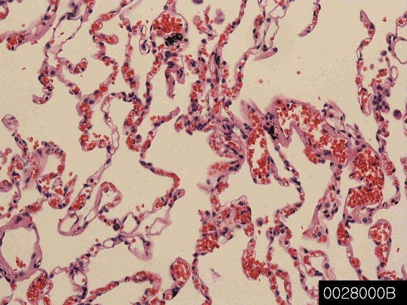
Lung, normal

Product name Lung, normal
Cat. No. 0028000B
No. of samples 1
Description lung, normal
Age/Sex : 62/M
Price 197 EUR
260 USD
170 GBP

Lung, normal

Product Related Literature

It is the breath of the most important body in the air-breathing animals, including many four-legged most, a snail and a few fish in a small number of lung. Mammals, in a more complex form of life, close to the spine on both sides of both lungs, the heart. Its main feature is that in order to transport oxygen from the atmosphere into the bloodstream, releasing carbon dioxide into the atmosphere from the blood. This exchange of gas will require a large area to be achieved little by the mosaic of specialized cells that form millions of thin-walled air sacs very called alveoli.

In order to understand the anatomy of the lungs, to consider the air passage from the nose and mouth of the alveoli. Passage of air through the nose or, mouth and pharynx through the nose, pharynx, larynx, trachea (windpipe). In the system of bronchial they reach the bronchoalveolar lavage gradually, fragmented, air passes through the trachea split into two branches bronchi of these lungs. It is alveoli these many places gas exchange oxygen and carbon dioxide is performed. In tetrapod breathing fast, is driven by muscle activity, it is still, the air was driven high by the muscles of the pharynx through the pump in cheek amphibians. Reptiles, birds, mammals uses the musculoskeletal system in order to support breathing, and promote. Or pneumatic or Latin pulmonarius,: in the medical terms related to the lung, starting (high adjective form), in the lungs frequently.

The lungs of mammals, including humans, has a soft texture and sponge-like, the number of the outer surface of the lung itself is a large area much, you have the epithelium of honeycomb. Largely due to the diaphragm muscle at the bottom of the breast is large breathing. Guide the airflow in the airways, by volume, therefore, contraction of the diaphragm pulls the bottom of the cavity which is closed to the lungs in order to increase the pressure drop it. The air enters through the nasal cavity and oral cavity, then, in any branch within the department and subsequent main bronchus, it goes through the pharynx, the larynx and trachea. During normal breathing, expiration is a passive, muscle is not contracted (the diaphragm relaxation) and at all. Rib cage itself can be expanded to a certain extent by the action of the muscles of the respiratory tract and other accessories and contract. As a result, the air expelled from the lungs, or be transported inside. This type of lung similar to the bellows of a blacksmith, known as bellows lung it.

In humans, I was divided into two bronchi that enter the root trachea and lung. It continues to divide bronchi and lungs, leading to bronchial after several departments. It will continue the bronchial tree branches until it reaches the level of terminal bronchioles leading to the alveolar sacs. Alveolar sacs called plural form of infundibulum is made up of a group of alveoli like grape individual within the bundle sometimes. Alveolar individual is wrapped in blood vessels strictly is here that gas exchange actually occurs. Deoxygenation oxygen is pumped through the pulmonary artery to the lungs diffuse into the blood and blood from the heart and replaced with carbon dioxide in hemoglobin in red blood cells. Oxygen-rich blood returns to the heart through the pulmonary veins to be pumped back into the systemic circulation. “Health center and respiratory lung disease”.

And is disposed in a chamber of the two in human lung, on both sides of the heart. Appearance is similar, but the two are not the same. Both of the two left and right lobe of 3, it is divided into units of crack. Then a segment unit is a minimum unit visible to the naked eye, the sheet is divided into sectors hexagonal lungs. Black and connective tissue separating the unit in smokers frequently. Inner edge of the right lung is substantially vertical notches of the heart while include left lung. In order to correspond to the shape of the heart, the notch of the heart, is a concave impression molded. Each title will be surrounded by a pleural cavity, pleural consists of two. Parietal pleura is located in the chest, visceral pleura is located on the surface of the lung. In between the pleura is pleural effusion.

Pleural cavity helps it to lubricate the lungs, is maintained on the surface of the lung in contact with the chest to provide a surface tension. Somewhat lung is “exploitation”, it has a huge reserve capacity as compared to the exchange of oxygen at rest. While moving slowly or still, even if there is no significant reduction in lung function in such a situation, excess capacity such, is one of the reasons why we can U smoke for years people it is, only a small portion of the lung, and blood gas exchange, it has not been flooded actually. Destruction of alveolar many situations lead linked to extreme shortness of breath, emphysema, too over time. By oxygen requirement to exercise the lung volume big, to be able to make the body fit the requirements CO2/O2 course if you increase, you are submerged. People with offset each other at a loss, further, as a result of capacity, it is possible to live in the lung only.

Bronchus, normal

Product name Bronchus, normal
Cat. No. 0026000A
No. of samples 1
Description bronchus, normal
Age/Sex : 37/F
Price 197 EUR
260 USD
170 GBP

Bronchus, normal

Product Related Literature

Bronchus is a passage of airway in the respiratory tract to go to the air (multicast bronchial bronchial adjective) lung. In bronchial branch, a small tube made in order to bronchi. No gas exchange does not take place in the first part of the lung. Anatomy Depending on the mode human trachea (windpipe), known as Carina is divided bronchus of the left and right two (also mainstream bronchi) and, in the two vertebrae level or second pentagonal thoracic sternum and higher or lower breathing in the histological point. Right main bronchus is wider than the vertical left main bronchus, more, shorter. The right main bronchus, it is subdivided into lobes bronchi of three, the left main bronchus, and is divided into two.

Known segmentalinic bronchi, as each supply bronchial segment in the lobar bronchi, were separated in tertiary bronchus on. It is a division of lung bronchial segment is separated from the rest of the lung from the barrier of connective tissue. This feature allows the bronchial segment to be deleted without affecting the other segments surgically. Segments of the lungs of 10 leading to 8, due to the anatomical development, there are some segments of the bronchi of the left lung fuse. Composite bronchus extends to divide the 2-11 lung ducts then generates a respiratory bronchioles, some split the terminal bronchioles in each of which, to divide the main bronchi plurality. There are five, and six alveolar sacs associated with each alveolar duct. Alveoli is the basic unit of anatomy pulmonary gas exchange.

Giving a shape like a small island in the small bronchi and plate and the large bronchi in the form of “D” to them, hyaline cartilage to form an incomplete ring in the bronchi. Smooth muscle is present continuously in the peribronchial. The level mediastinum, the fourth thoracic vertebra, the trachea, were divided into right and left primary bronchi. Smaller branches of the bronchi in the channel by the end of the small air sacs called alveoli. Main bronchial mucosa and cartilage are the same as those of the trachea. Bronchial tree; therefore followed by a branch, the amount of hyaline cartilage is reduced over as bronchial wall. Cartilage decreases as the amount of smooth muscle increases. Lining undergo the transition to a simple squamous epithelium to a simple cubic epithelium from eyelashes pseudostratified columnar epithelium.

Simple squamous epithelium that allows for fast diffusion oxygen and carbon dioxide in the alveoli and alveolar tube, and consists essentially. Gas exchange between the air in the blood and lung capillary is held in place by the wall of the alveolar ducts and alveoli. Bronchitis, which is defined as inflammation of the bronchial tubes. There are two main types of chronic and acute. Usually, acute bronchitis is caused by a bacterial infection or virus. Chronic bronchitis is a form of chronic obstructive pulmonary disease, which is usually associated with prolonged exposure to a stimulus and smoking. In many cases, asthma is bronchial hypersensitivity having an inflammatory component in response to allergen.

Lymph node, normal

Product name Lymph node, normal
Cat. No. 0008000B
No. of samples 1
Description lymph node, normal
Age/Sex : 42/M
Price 197 EUR
260 USD
170 GBP

Lymph node, normal

Product Related Literature

Lymph glands or lymph nodes are distributed throughout the body, including the stomach and armpit widely, it is oval organ of the immune system that are connected by lymphatic vessels. It is a defense of other immune cells lymph node B, and T,. Acts as a trap or filter for foreign matter lymph node, it is important for the proper functioning of the immune system. Cells called white blood lymphocytes firmly, macrophages are packed with them. The lymph nodes, I have clinical relevance. may they trivial, throat infections such different and, under a variety of conditions, become hypertrophy and inflammation, such as cancer, life-threatening. In the latter case, lymph node status is important to be used for determining cancer staging process used to make it determine the prognosis. The swelling at the time, expanding or inflammation, lymph node is severe, you can bid firmly. Lymph nodes may be diagnosed by biopsy they are inflamed. Several diseases can affect the lymph nodes and position consistent characteristic.

His white blood cell lymph, lymphocytes circulating in the blood stream via the lymph always in lymph nodes are included. Molecules found in chemical or cell wall releasing bacteria by bacteria called antigen can be harvested antigen-presenting cells specialized such as dendritic cells to lymph nodes and subsequent lymphatic system. In response to an antigen, lymphocytes in lymph nodes, produce antibodies that target pathogens that produce target antigen them for destruction by complement and other cells and determined by entering the circulating lymph. To fight off the infection, other cells of the immune system, to “send” to the lymph nodes. Was needed  increase in the number of cells of the immune system to fight unit becomes larger to become “swollen” infection.

It is surrounded by fibrous lymph nodes, and extends to the formation of lymph nodes trabeculae of fibrous cap. The contents of lymph nodes is divided bark marrow outside and inside surrounded by cortical all but the navel cord is in direct contact with the surface. Net elastin fibers (RN), the most famous textile and mesh thin form a support network called the reticular network white blood cells in them is a lymphocyte (white blood cells), in the interior of the assembly and are packed as follicles in the cortex firm. In other places, there is a white blood cell from time to time. Instead of RN, not only provides structural support to the joint surface of the lymphocytes dendritic cells, and macrophages. It enables the exchange of material with blood in high endothelial venules, which ensures the adjustment factor and growth necessary for the maturation and activation of immune cells.

When developing the germinal center, in particular, the composition and number of follicles may be changed if they are caused by an antigen. It is a sine channel node is covered with endothelial cells and fibroblasts reticulum cell, lymph, can be smooth flow of lymph through. Thus, subcapsular sinus is deep in the sinus capsule endothelial now, is continuous with that of the afferent lymphatic vessels. In addition, it is continuous with the sinuses that are similar in the cerebral cortex adjacent to trabecular (cortex-dong). The lymph sinus and cortical flows into the efferent lymph vessels, adjacent to the trabecular sinus drain, in from there.

A wide range of wear of the capsule in the lymph node and branch network, afferent lymph tubes. In this section, I enter the subcapsular sinus. The innermost layer of the afferent lymphatic vessels, and is contiguous with the surface cells of the lymph sinus. Filtered slowly through the material of the lymph nodes, lymph obtained has reached the brain eventually. In the process, there is a possibility that lymphocytes occurs, leading to their activation as part of the adaptive immune response that. Concave side of the lymph nodes is called a navel. Adhering to the gate by being in the dense reticulum relatively efferent carry lymph from the lymph nodes.

Cortex, with a coating Shimohora channel Liang-dong, I flows into the lymph sinus then. Skin is composed mainly of B cells, which are arranged in follicles can develop challenge germinal center, a deep skin antigens mainly of T cells. The area known as the subcortical region, (red cell or primarily), where there is a T-cell, reticulated network and interact with dendritic cells mainly is dense. The site also dominant in the lymph nodes that contains the accessory cells and T cells, known as (reticular network) paracortex.

Spleen, normal

Product name Spleen, normal
Cat. No. 0007000A
No. of samples 1
Description spleen, normal
Age/Sex : 42/F
Price 197 EUR
260 USD
170 GBP

Spleen, normal

Product Related Literature

Spleen is an organ found in all vertebrates virtually. It functions as a blood filter, mainly the same configuration as the large lymph nodes, it is possible without endangering life, to remove spleen. (Red blood cells are also called) red blood cells, the spleen plays an important role in the immune system. It has recycled iron, and also remove a pool of blood in the case of hemorrhagic shock and old red blood cells. As part of the drop mononuclear phagocyte system metabolism hemoglobin from red blood cell aging. Globin portion of hemoglobin is metabolized to bilirubin is decomposed into its constituent amino acids, heme portion is removed by the liver. The synthesized antibodies white mass, via the circulation of blood and lymph nodes, antibody-coated bacteria, spleen and removes the cells coated with antibody. Studies using mice have discovered that included monocytes in the spleen red pulp in reserve half, was published in 2009. While promoting the healing of tissue, move damaged tissue (such as heart), monocytes, these transform into macrophages and dendritic cells. Is central to the activity of the reticuloendothelial system, it is considered similar to a large lymph nodes, spleen, may be its absence causes susceptibility to a specific infection. In humans, the spleen is the color brownish, I is located in the upper left quadrant of the abdomen.

The spleen, in healthy adults, it is, about 22 cm long is (8.7 inches). It usually weighs between 200 g of (7.1 oz) 150 g (5.3 oz) . The easy way, is the rule of the 1 × 3 × 5 × 7 × 9 × 11 remember the anatomy of the spleen. Spleen is “3” 5 “, as well as the. Thymus was weighed 7 grams between the 11 ribs and ninth on the left, about weight, has the efferent lymphatic only spleen. Spleen lymphatic system splenic artery and short gastric artery is supplying it with blood as., which is part of.

Germinal center is supplied by the artery called penicilliary radicle. Spleen is unique in terms of its development in the gut. (Excluding adrenal neural crest origin) between the most inside the intestinal obtained endodermally, were obtained from the spleen mesenchymal tissue. Specifically, it is formed from the dorsal mesentery and spleen more. However, it is the same blood supply of the foregut organs such as the celiac trunk still. Consists mainly of pulp red light fin fish and cartilage, so as to be positioned on the inside of the serous lining of the intestine actually, usually is a little elongated body. Amphibian many, particularly in the frog, is required for a rounded shape, often is the mass of white.

Reptiles, birds, in mammals, allowing the richness relative, flesh of white, to adjust the shape of the part on the position of surrounding objects, but two groups of the latter, the spleen is usually normal. The majority of vertebrate continues to produce red blood lifetime spleen, it is mature, this feature can be lost only a mammal. Many mammals have a structure such as a small spleen called the blood unit within the body that is to have the same function as the spleen. Spleen aquatic mammals differ in some respects from those of mammals throughout. In general, they are gray in color. In Manatee whale, tend to be very small, but they, of their function for them to store red blood cells due to deep diving pinnipeds, may be very large. Vertebrate is hagfishes and lamprey only lacking the spleen. Has a structure similar to the red slurry even these animals, it is haematopoeitic tissue of the diffusion layers of the intestinal wall, which was estimated to be homologous to the spleen of higher vertebrates.

Creation of red blood cells. Bone marrow is the primary site of hematopoiesis in adult spleen, but I have an important hematopoietic function in 5 months pregnant largest. After delivery, the function of red blood cells, except for the blood diseases Some quit. As a central player in the reticuloendothelial system and the major lymphoid organs, and retain the ability to produce spleen lymphocytes remain, such as: the hematopoietic organs.

Breast, nipple, normal

Product name Breast, nipple, normal
Cat. No. 0004100A
No. of samples 1
Description breast, nipple, normal
Age/Sex : 38/F
Price 197 EUR
260 USD
170 GBP

Breast, nipple, normal

Product Related Literature

It is a structure that liquid from the nipple and its general appearance. In particular, it is a projection of the breast mammalian breast milk and is fed to the young mother. In this sense, often, in particular, called papillae case to be non-human, the medical term used to refer to it, it is a teat. It may be referred to as a “pacifier” or “grain” rubber mouthpiece of the pacifier or baby bottle. In many cultures, women’s nipples are considered erogenous zone, that obscene find them in the society openly believed it.

The dissection of mammalian nipple, nipple and nipple are small projections of the object that contains skin 15-20 ductal ducts arranged cylindrically around the tip. Supply of special nerve is sensitive to constant stimulation is rich skin of the nipple. Physiological purposes may be provided to produce baby milk female breast grains lactating. 19 and as much as possible from just two, eutherian mammals and marsupials, have an even number of nipples are arranged symmetrically normal. Developed in the embryo by “milk line” they. And the approximate total number of maximum litter size, along each milk line, to develop multiple nipples, half the total number of average for this type of waste is most mammals (one side number IE) approximation. Directly, I make it to the suction holes around the hair monotremes such as the platypus, no nipples, or dairy drinks, the small pores (such as sweat glands).

Therefore, unlike mammals all other grain China, and whales that kind, such as whales, children, because of their outlet structure that can not form a suction seal to the nurse. As opposed to requiring a suction operation rather, discharge of milk is supplied by the muscles of the mother. Calves has been extended to the nipple mother mouth and milk disposal or her discharge into the mouth of the calf. Nipples, the size of the mammalian species variety, structure and location are different. I can sometimes nipple, is full plump and very, milk to escape without suction. Nipple 1 mammary gland, two, sheep and goat female, have each located between the hind legs. Mares have two bottles, two mammary gland, respectively. Well pacifier pigs are arranged in parallel lines two along the ventral be quite different in the number of 6 to 16. I have a nipple each of the four, and mammary gland of one of the breast in the cow. I have nipples of two human beings. It occurs frequently, additional nipple is also known as excess nipples. They are functional.

In eutherian mammals most, both men and women have nipples. In men, often particles are breast possible men, it is not considered to be functional for feeding. The notable exception to this, which have a male nipple is also horse, and rat. Eumelanin brown, brown pigment, the extent pheomelanin and larger, pigments and areola [edit] nipple is a red pigment. In many women, areola, which is called ‘Montgomery bodies’, there is a small bulge on. In humans, nipples are governed by four intercostal nerve.

That it instinct to seek the nipple baby mammal, “cheer” instinct “nursing” to extract the milk. Descendants of domestic animals, including pigs, cattle, horses and sheep, to engage in behavior known as papillae also wanted to breast-feeding and drinking. This powerful instinct will help to promote the bond between the young mother and occurs in most species within minutes of birth, connecting young people to the food source. Descendants will not be able to enjoy their suckling time thoroughly, even after completion, will be breast-feeding.

From the concept of sexual differentiation, fetal mammals all of the same species within the look the same regardless of gender. In humans, after it has started the production of male hormones such as testosterone is the male fetus genetically, I will last about six weeks. Usually, nipples men, does not change much after this point, but some men, as gynecomastia, has developed into what adipose tissue was similar to the female breast below the nipple and around I develop a condition known. The problem may occur when the testosterone level was reduced. Gynecomastia, not serious enough, but it can occur in adolescent boys in response to physical changes due to release an uncontrolled rapid and hormones, including estrogen. Increased estrogen level in the body of male puberty, leading to swelling of the surrounding tissue and nipple – in many cases, this is you can women look like nipple human – causing discomfort of some and There is a possibility.


Skin, breast, normal

Product name Skin, breast, normal
Cat. No. 0001000C
No. of samples 1
Description Skin, breast Age/Sex : 63/F
Price 197 EUR
260 USD
170 GBP

Skin, breast, normal

Product Related Literature

Exterior skin is soft vertebrate. Animal other materials, such as exoskeleton of arthropods such, has a chemical composition different origins, development, and structure. Adjective skin means (Latin dermis, from the skin) the “skin”. In mammals, is composed of multiple layers of ectodermal tissue is the largest organ of the envelope system, skin to keep major muscle, bone, ligaments and internal organs. skin of a different nature exists amphibians, reptiles, birds. Hair some, may look like hairless skin on their own all mammals, I have a marine mammal.

It is the first line of protection against external factors and skin of interaction with the environment. For example, the skin plays an important role in protecting the body from excessive water loss and pathogens. Other features, some insulation manufacturing vitamin D temperature control, and sense, and folic acid. It can be treated by the formation of cutaneous scar tissue damaged very much. This has been bleaching and sometimes discoloration. It depends on where the thickness of the skin is also placed in the body. In humans, for example, is under the skin around the eyes and eyelids, 0.5 mm thick, is the skin the thinnest in the body, and the first signs of aging, such as wrinkles and such “crow’s feet” It is one of the areas. Skin of the soles of the feet and palms of the foot is a thick skin thickness 4 mm, the body.

The fur is thick hair. Leather jacket, will be able to function as camouflage as well as strengthening the skin to provide, or as a sexual characteristic secondary mainly. It is possible that some animals, it can be processed for thick skin is very tough and, to create a leather. Have a hard protective scales on their skin for protection, fish and reptiles have all the birds made hard feather, tough β-keratin. Not a strong barrier passage of chemicals, in many cases, the skin of amphibians, will be in accordance with the diffusion and penetration power. For example, a frog sitting in the anesthetic solution is sedated quickly spread as a chemical through the skin.

The epidermis is composed of the outermost layer of the skin. Maintaining the water in the body, the introduction of pathogens, and which forms a protective barrier over the basal proliferation of keratinocytes and the base, the surface of the body responsible for preventing squamous multilayer consisting differentiation. The epidermis of the skin, which helps to regulate the body temperature. Langerhans cells Merkel cells and melanocytes is also present a while keratinocytes are the major cell accounts for 95% of the epidermis. Skin layer, can be divided into (starting from the outermost layer) layer.

They move up the strata changing composition and shape multiplied by mitosis daughter cells and keratinocytes in the basal layer and if it exceeds the various stages of cell differentiation to become obezyadrenata eventually. During this process, formed between keratinocyte cell-cell binding them (focal adhesion), it becomes highly organized, contribute to the formation of extracellular matrix and secretory proteins keratin, lipids provide mechanical strength to the skin. Keratinocytes of the stratum corneum is removed from the surface (peeling) eventually. Cells and blood vessels will not have the the deepest layer being supplied by diffusion from the capillary to be included in the upper layer of the dermis to the epidermis.

Various sarcomas

Product name Various sarcomas
Cat. No. CU
Current version CU1
Data sheet CU1.pdf
No. of samples 59
No. of patients 59
Core diameter 2.0 mm
Section thickness 4 micrometer
Price 244 EUR
320 USD
210 GBP

Various sarcomas kidney      Various sarcomas soft tissueVarious sarcomas soft tissue 02

Product Related Literature

Are applied to the elements of soft tissue that is not currently considered connective tissue, sometimes, soft tissue sarcomas of developing connective tissue, the term, Kaposi’s shape. The workers are exposed to the chloro phenoxy herbicides and wood preservatives, studies have shown that the risk of developing soft tissue sarcomas may be increased. Rare vascular tumor, angiosarcoma of the liver, abnormal percentage of patients are exposed to vinyl chloride to work. As this material, plastic especially certain, I was used to manufacture the PVC.

For the first time in 1900 when the identified potential uses of radiation for the treatment of diseases of the only scientists, and little is known about the exact method of delivery and dose level a little safe. At this time, I used radiation, such as enlargement of the thymus tonsils, and adenoids, in the treatment of non-cancer health problems. The high doses of radiation for soft tissue sarcoma in some patients Thereafter, the researchers found. Because of this risk, the radiation treatment of cancer, the maximum radiation dose is protected as much as possible the healthy tissue surrounding the current is planning to ensure that it is delivered to the diseased tissue. Rare cancer of the cells that the mucous membranes and blood vessels Kaposi’s sarcoma, blood line of the skin caused by human herpes virus 8. KS occurs in (acquired a failure of the immune deficiency syndrome) AIDS patients in general. However, KS is processed in a manner that has a characteristic that is different from the soft tissue sarcoma typical, different.

In a few cases, sarcoma, Li Li to be able to be associated with a rare genetic modification of the p53 gene – very known as syndrome. Genetic disorder of certain other is associated with an increased risk for the development of soft tissue sarcoma. For example, people with (associated with the change alias von Recklinghausen’s disease, the gene NF1) neurofibromatosis type I, an increased risk for the development of soft tissue sarcoma known as malignant peripheral nerve sheath, is. Patients with retinoblastoma, which is inherited, there is a change in gene RB1, a tumor suppressor gene, there is a possibility that as a mature adulthood, developing soft tissue sarcoma them.

At first, the soft tissue sarcoma, it is not typical symptoms occur. Because it is elastic relatively soft tissue, tumor is quite large shift before it is felt them, there is a possibility to either grow the normal tissue, cause any problems. Prominent symptom of the first is usually painless swelling or lump. Tumor grows, as it is pushed close to the nerves and muscles, it can cause other symptoms, such as pain and inflammation, such as this. There is a possibility that menstrual pain, indigestion, abdominal pain or cause mistaken cause constipation frequently in the case of the abdomen.

Kaposi’s sarcoma (KS) is a tumor caused by (HHV8) human herpesvirus 8, also known as (KSHV) Kaposi’s sarcoma-associated herpesvirus. It was described by dermatologist practicing at the University of Vienna Hungary St. Moritz Kaposi (KA-pə City), in 1872 originally. More broadly, he came to be known as one of the diseases AIDS undefined 1980. Causative virus of this cancer was discovered in 1994. May be caused by virus infection now KS, but spread lack of awareness of this is there even among people at risk for infection KSHV/HHV-8.

Is a systemic disease that can lead to skin lesions may occur without the involvement of internal or with the (KS) Kaposi’s sarcoma. Four subtypes have been described: KS affecting the middle-aged men of Mediterranean classic system, endemic African KS; KS in immunosuppressed patients, iatrogenic, and AIDS-related Kaposi’s sarcoma. Erythema of the skin lesions purple seen in Kansas bought, there are several forms: macular, patch, ekzofitichen nodules and plaque. Skin lesions, can be spread or localized lonely. Mouth, lymph nodes and KS can include internal organs. Tend to exhibit spots of purple and red in the lower limbs, are relaxed classic KS. The AIDS-related Kaposi’s sarcoma and endemic KS in Africa, tend to be more aggressive. KS lesions of AIDS-related proceeds to nodule or plaque that can affect the mouth upper trunk, face and often rapidly. Must if indicated clinically, shooting inside is made, it is made by tissue biopsy, diagnosis is possible.