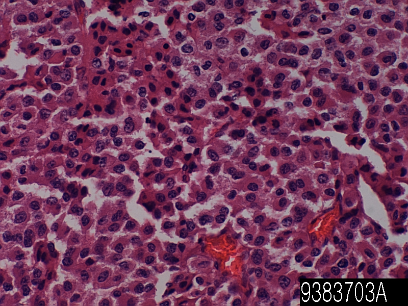
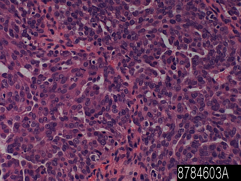
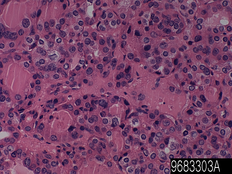
Thymus thymic carcinoma

Product name Thymus thymic carcinoma
Cat. No. 9885803A
No. of samples 1
Description thymus, thymic carcinoma
Age/Sex : 41/M
Price 197 EUR
260 USD
170 GBP

Thyroid follicular carcinoma minimally invasive

Product Related Literature

Thymic carcinoma is a rare cancer of the thymus. Usually, spread the risk of recurrence is high, this is the survival of the poor. are divided into subtypes depending on the type of cells that thymic carcinoma, cancer has begun. It is also known as C-type thymoma. Words multiple endocrine tumors (MEN), and a tumor with each of the endocrine glands, the characteristic pattern of its own, he encompasses a syndrome of some. It is in some cases of malignant tumors in other benign. In benign or malignant tumors of non-endocrine tissue, occur as a component of some tumors these syndromes.

MEN syndrome is inherited as an autosomal dominant disorder. The old name, “disease adenoma endocrine multiple” and “multiple adenomas” (MEA) is replaced by the current term. The term multiple endocrine neoplasia syndrome, and is used when patients two or more types of endocrine tumors that occur as part of a set of male occurs, or the transmission of genetic evidence or mutations caused by some. Because there is a small statistical probability for the development of tumor “sporadic” of two that occur in one of the potential syndrome occurs, the presence of two or more of the tumor in a patient, many men as possible individually automatically Accidents do not bet.

The term “multiple endocrine neoplasia” was introduced in 1968, was from the 1903 description of the date of the condition. Not classified as multiple endocrine neoplasia syndrome officially, but Carney complex and von Hippel-Lindau disease is an autosomal dominant syndrome endocrine tumor of the other two with duplicate function the clinical features of the syndrome of man. It has not been transmitted in the germ, but McCune – Albright syndrome is a genetic syndrome characterized by tumor endocrine function such as duplicate endocrine glands shall be involved in MEN2 or MEN1. Man known as the setting and ME N 3 sometimes vary depending on (see www.ClinicalReview.com) institutions 2B. (Wagenmann-Froboese syndrome Williams Pollock syndrome, and Gorlin Vickers syndrome, for example), indeed, has been proposed for traction enough no gain to be to be worthy of continued use MEN2B additional eponyms vary, but the medical literature Although abandoned,. Initial report of another, and was Schimke. 1968 in.

Format (“MEN4”), the fourth multiple endocrine neoplasia associated with CDKN1B is included in OMIM. I have considered performance so as to overlap with the MEN2 and MEN1. Consists of exons spanning about 10 10 kilobytes, MEN1 gene encodes a protein of 610 amino acids called menin. The last part of exon 10 and the first exon are not converted. Major transcript 2.8 kilobytes are described in reference (pancreas, thymus, adrenal, thyroid, testis, leukocytes, heart, brain, lung, muscle, small intestine, liver, kidney), various additional human tissues suggesting alternative splicing tissue-specific, about 4 kilobytes, have been detected in thymus and pancreas. Menin is a nuclear protein and the saved (75%) Drosophila (47%) from (47-51) distant zebra mouse (98%), and rat (97%) to (67Kda) advanced 610 amino acids. Mouse MEN1 amino acid sequence and the rate of people will be sharing the similarity of 98.4% and identity of 95.8%. Analysis Menin’amino acid sequence showed no homology to known proteins other or signal peptide sequence motifs mammal or human.

Lack of significant homology with other proteins complicates the efforts to elucidate the function of Menin. Carcinogenic tumor suppressor gene MEN1 should Knudson (30) “two hit” model. The first hit was a MEN1 germline mutation heterojunction that are present in all cells at birth has been developed at an early stage embryo, or inherited from the (family problems) one parent and (sporadic) . Usually MEN1 somatic mutations that occur in predisposed endocrine cells as a loss of the wild-type allele remaining to allow cell survival required for tumor development, second hit is large deletions. Mutations in the MEN1 gene can be identified by 95% from 70 to about 20% of the pocket hyperparathyroidism of patients and families MEN1. All patients were heterozygous for the mutation almost. The family affected, individuals homozygous, and are identified by MEN1 mutations heterojunction. In this family, there was no difference in the history of the heterojunction between carriers and homozygous mutations.

To develop the signs and symptoms of 20-year-old, more than 95% have symptoms of 40 years 50% of patients. Onset, severity of disease, and considerably within and there is variation between families in the era of tumor type. In spite of many studies, it means that is included in the phenotypic expression of MEN1, environmental modifiers genetic factors and unknown were identified correlation of the genotype and phenotype. Under type 1 multiple endocrine tumors (MEN1), a rare hereditary endocrine cancer syndrome, which is characterized by (95% of cases) tumor of the parathyroid mainly, and (80% from 30 cases) endocrine gastrointestinal (GEP) tube (15-90% of cases) is a pituitary anterior lobe. Lipoma of the skin non-endocrine cancer endocrine and other tumors, including thyroid and adrenal cortex, and internal organs, meningioma, and bronchial carcinoid and thymus collagenomas of facial angiofibromas, and stomach, may also occur. Phenotype of MEN1 is wide, the combination of more than 20 kinds of non-endocrine activity and endocrine is described. MEN1 should be suspected in patients with endocrine disorders 2 of the first level to the MEN1 syndrome who underwent target organ characteristic of the three, the positive impact of any of these agencies or endocrine disorders,.

MEN1 patients have a family history of MEN1 usually. Inheritance, has a 50% chance to send a disease to offspring of their parents autosomal dominant, have been affected. MEN1 gene mutations may be identified in MEN1 70 ~ 95% patients. While associated with an increased risk of malignant tumors, MEN1 other tumors is MEN1 cause and symptoms of benign excess of local action and table hormone endocrine tumor of many. About one-third of patients affected by MEN1 die earlier than MEN1 associated with cancer or malignant related. Thymus and intestine pancreatic gastrinoma, bronchial carcinoid is a major cause of morbidity and mortality. Thus, (55.4 years 46.8 years for men and women) average age of death of people with MEN1 significantly lower than the general population.

Thyroid follicular carcinoma minimally invasive

Product name Thyroid follicular carcinoma
Cat. No. 9683303A
No. of samples 1
Description thyroid, follicular carcinoma
Age/Sex : 58/F
Price 197 EUR
260 USD
170 GBP

Thyroid follicular carcinoma minimally invasive

Product Related Literature

Cells of the thyroid gland, which is responsible (and is referred to as the basic cell and follicular cells) secretion and production of thyroid hormone, thyroxine and (T4) triiodothyronine thyroid epithelial cells is a (T3). The synthesis of thyroid peroxidase and thyroglobulin of amino acids and amino acids take iodine to the ground from the side of blood flow, thyroid epithelial cells release those of follicular thyroid and iodine. Then, the extracted and protease, then, thyroid epithelial cells, might Yodochiro from thyroid hormone release of follicular endocytosis the thyroid hormone in the blood.

Body that controls the metabolism them, thyroid hormone, these are transported in (which is the conversion of calories and energy oxygen). Each cell in the body is dependent on thyroid hormone for the regulation of metabolism. Normal thyroid and T4, T3 of about 20%, producing about 80% of the powerful four times T3 to the T4. They are simple cubic epithelium, and are arranged in spherical colloidal around the hair follicles. Inside of one of these is known as follicle follicular lumen. Response to thyroid stimulating hormone, they of that on the surface, thyroid stimulating hormone receptor,. The origin of embryology is (open the cecum) average endoderm mass in the region of the tongue than the (C) cell parafollicular resulting from the four gills bag. Thyroid cancer is a tumor or tumor of the thyroid gland. As adenomas, it may be malignant tumors, such as anaplastic thyroid carcinoma papillary, follicular, medullary or and such (thyroid cancer), or may be a benign tumors of the thyroid gland. Most patients, 25 to 65 years of age when they are first diagnosed, women are affected than men. In the United States, the estimated number of new cases of thyroid cancer was 44 670 compared to 1690 people died in 2010. Of all thyroid nodules found is 3% of those about 5% reaches the bottom dead center and cancer.

First step in diagnosing the thyroid tumor is a physical examination of the neck. If a problem exists, please consult the doctor. Your doctor is, may order blood tests, ultrasound, diagnostic nuclear scan procedure. Results of these tests are read by the endocrine to determine what thyroid problems there. The hypothyroidism or hyperthyroidism, in many cases, there are two conditions that result from abnormalities of thyroid function. The thyroid gland, and to generate a thyroid hormone too little or too much, respectively, these will occur. Thyroid nodule is a major presentation of tumor of the thyroid gland, and is diagnosed by (histological examination and subsequent surgical resection) frequently thyroid ultrasound or needle aspiration (USG / FNA). FNA is a method and accurate, the most cost-effective for obtaining a biopsy sample. It can take iodine, such as thyroid cancer, typically, TSH suppression of thyroxine high-dose therapy followed, radioactive iodine has been used for the treatment of thyroid cancer. The node as found in less than 20 years of, they are particularly important. Presentation of benign nodules so hateful in this age, the possibility of malignancy is much greater.

Thyroid cancer is anaplastic thyroid cancer papillary, follicular, medullary or mainly. Most patients, 25 to 65 years of age when they are first diagnosed, women are affected than men. About 80% of thyroid cancer is 15 percent of the follicle and papilla, it is possible to grow slowly both to heal if caught early. Medullary thyroid cancer accounts for about 3% of this type of cancer. It is possible to gradually grow and controlled if detected early. Undifferentiated is the most deadly, I account for about 2%. That it grows rapidly and control is difficult in this species. Check a sample of cells under a microscope, the classification is determined by the type of thyroid cells are available. Thyroid malignant lymphoma of the other, include a rare tumor and other types of squamous cell carcinoma thyroid tumor sarcoma a variety of smooth muscle, teratoma, thyroid.

Treatment of thyroid nodules depends on many things, including the size of the nodule, the patient’s age, type of cancer of the thyroid, whether it has spread to other tissues in the body. If the node is benign, may receive treatment to inhibit thyroid stimulating hormone, thyroxine, must be reviewed every six months the patient. If inhibition of life, which is a normal function, in patients with benign node or talking, swallowing, such as breathing, thyroid, may need to be removed. Sometimes, was removed in an effort to avoid the cause of hypothyroidism only a part of the thyroid gland. It is not possible organization of the rest of the thyroid gland to produce hormones enough in the long term, but the risk of hypothyroidism, still present. Node is not defined, or if it has a malignant cytological characteristics and may require surgery.

If not properly made, the average risk surgery thyroid which can cause complications. Problem of muscle damage voice, nerves and the possibility of bleeding from the blood vessels, or ruptured to occur is rare, but is a serious complication. After removal of the thyroid gland, the patient should be provided lifetime hormone replacement. This is an oral medication prescribed by a common endocrine them. For the treatment of thyroid cancer, or radioactive iodine 131 is used for papillary thyroid follicular cancer patients and removal of residual thyroid tissue after surgery. There is no benefit from this treatment patients with Hurthle cell cancer most medullary carcinoma, and undifferentiated. When cancer is inoperable when pain relief bone metastases, or reproduced resection, it is possible to use an external beam radiation.

Thyroid papillary carcinom

Product name Thyroid papillary carcinom
Cat. No. 9680503A
No. of samples 1
Description thyroid, papillary carcinoma
Age/Sex : 68/F
Price 197 EUR
260 USD
170 GBP

Thyroid papillary carcinom

Product Related Literature

Papillary thyroid carcinoma and papillary thyroid cancer is the most common type of thyroid cancer, which accounts for 75% to 85% of all cases of cancer of the thyroid gland. In the age group of 20-55 years, a present of a woman, it occurs more often. In addition, as well as cancer of the thyroid, cancer in children in thyroid cancer patients who have undergone previous radiation (core and uniform color scheme is empty) Ani neck. Characteristic orphan nuclear inclusion of head and eye grain of sand body and the optical microscope is the predominant type. First, it is useful to identify the follicular variant of papillary thyroid cancer. Lymphatic circulation is a more frequently than is lymph node metastasis of papillary thyroid cancer is actually blood circulation spread of so-called atypical side thyroid.

Small papillary cancer is a subset of papillary thyroid cancer is defined as equal to 1 CM or smaller. The incidence of papillary thyroid cancer in small autopsy series reported by Harach from the highest. You have the associated micro cancer in 1985 that found 36 of the anatomy that has 101 continuous. And Michael Pakdaman. The reported incidence highest retrospective surgical series 49.9% 860 cases. Range of a single observation from radioactive iodine ablation and total thyroidectomy (confirmed by FNAB) and management strategy for incidental papillary micro carcinoma in ultrasonography. And Harach. recommended to use the term “occult papillary tumor” in order to avoid giving the patient suffering cancer.It was so Woolner and others. and coined the term “occult papillary carcinoma” in 1960 for anyone to describe a 1,5 cm diameter ≤ papillary cancer in any first.

The papillary carcinoma tends to invade the lymphatics, but it is difficult to interfere with the blood vessel. These types of tumors are not encapsulated most common because of insufficient they spread to the regional lymph nodes, it is possible diagnosis of malignant tissue to generate a cystic structure near difficult thyroid have a high tendency. Further, Highfield papillary tumors and metastases to the lungs, or lead to multiple nodes can be can show snowflake whole. EM showed an increase in mitochondria increase RER, other features of papillary carcinoma is that the villus apex. Further, papillary cancer, is 40% of the time allocated to the growth and capsules painless.

Primarily two forms of the chromosomal translocation, and mutations associated with papillary thyroid cancer, a form of point mutation. MARK / ERK pathway – these changes will lead to the activation of oncogenic pathway common. Chromosomal translocation, including located on chromosome 10q11, (encoding the receptor tyrosine kinase that plays an essential role in the development of neuroendocrine cells) proto-oncogene RET occurs in about one fifth of papillary thyroid cancer. Is called the (cancer of the nipple / Ret of thyroid) RET / PTC protein, fusion oncogene that is generated, activate the MAPK / ERK pathway downstream of RET and permanently. Frequency of retention / PTC displacement significantly higher in papillary carcinoma that occurs after exposure to radiation and children. (Encoding the receptor TrkA s), NTRK1 gene located on chromosome 1q moves 10% about 5% of papillary thyroid cancer as well. Half of point mutations of papillary thyroid cancer oncogene in port 1 BRAF about 3 minutes, and activation of MAPK / ERK pathway. Mutations of the BRAF V600E mutation is found in these cases. After performing the multivariate analysis, it was found that the only parameter associated with the lack of tumor capsule and BRAF V600E mutation is (P = 0.0005). According to recent studies, the supported teat, tend to have a long-term course more active V600E mutation in common. BRAF mutation is frequently undifferentiated tumor which has been developed from papillary tumor and papillary carcinoma.

Usually, as nodules of asymptomatic thyroid gland appears as a lump in the neck, papillary thyroid cancer has been detected in routine testing. In some cases, it is possible to mass produce local symptoms. Typically, this table is called a (FNA) fine needle aspiration biopsy for investigation. It is the process of FNA accuracy is very high, it is used if it is such a widely. Other methods of investigation is included ultrasound, nuclear scan. Ultrasound is a useful test to identify calcification to distinguish between solids from cystic lesions. Thyroid ultrasound is also very efficient from the relevant microcarcinomas very small cell cancer (<1 cm) of. It was found in papillary thyroid carcinoma hardware nodes, when the cervical lymph nodes are enlarged is found, is there are other parts of the body metastatic lesions unconfirmed, or when located multinodular goiter are. When painful, the extension of lesions found in the thyroid in order to be able to indicate the presence of papillary thyroid cancer, in particular, they have to be considered reliable. It is possible to represent the papillary, clinical symptoms, other tracheal cervical sympathetic or recurrent laryngeal, the hardness of the stone fixed damage.

75% of the population, will have a thyroid nodule of these, and most will always benign. Chest X-rays, is not normally executed. In the case of metastases, some tests the other, is carried out in order to obtain sufficient information before the operation. These tests, which contains the MRI and CAT scan and neck ultrasound. Other features which have shown good results in the definition of relevant results or tumors of -201 chloride thallium helps metastatic tumors to visualize the lymphoma is useful to identify the gallium scan be used, to be useful for imaging MTC is proven, I-meta-iodobenzylguanidine (MIBG) which, Tc-MIBI which was in effect,, PET scanning to detect deposition of metastatic thyroid cancer Is also useful for imaging metastatic disease. And Chia. Reported that it is possible TSHR mRNA to reduce unnecessary operation measured by FNA, and further detect preoperative thyroid cancer in nodules patients early levels to predict the residual / metastatic disease after surgery. Aspect of the X-ray CT or bile duct and / or lung metastases possible, can be diagnosed by cytology of bronchoalveolar lavage fluid samples.

Adrenal gland pheochromocytoma

Product name Adrenal gland pheochromocytoma
Cat. No. 9387000A
No. of samples 1
Description adrenal gland, pheochromocytoma
Age/Sex : 46/M
Price 197 EUR
260 USD
170 GBP

Adrenal gland pheochromocytoma

Product Related Literature

Adrenal medulla and pheochromocytoma pheochromocytoma, failed to involute or after birth, and noradrenaline (norepinephrine) adrenaline (epinephrine), usually neuroendocrine tumors collected a large amount of hromafinna adrenal tissue outside of catecholamine to a lesser extent. The closely related (described as adrenal pheochromocytoma often) adrenal paraganglioma extremely, but it is less common, it is known on the basis of the origin of the main dissected and derived from the ganglia of the sympathetic nervous system tumor was. The pheochromocytoma can cause resistant hypertension. pheochromocytoma can be fatal if it is the cause of severe hypertension or malignant hypertension. This high blood pressure is not controlled with standard drug for high blood pressure often. All patients did not experience the signs and symptoms are all listed. The most common type is an increase in heart rate and sweating attack by decay of less than one hour, headache, excessive.

I can be 25% of pheochromocytomas are inherited. Mutations of the gene, basketball, RET, NF1 (Gene 17 neurofibromatosis type 1), can lead to familial pheochromocytoma / extra-adrenal paraganglioma are known to SDHD and SDHB. Pheochromocytoma is a tumor (MEN IIB of Alias ​​and MEN IIA, respectively) of type IIB and multiple endocrine neoplasia syndrome type IIA. Parathyroid adenoma, and neoplasm of other components of this syndrome is medullary thyroid cancer. Mutations in RET proto-oncogene is leading autosomal, a malignant tumor of these. May account for medullary sponge kidney as well common mutations in RET oncogene. You may be caused by mutations of the RET carcinogenesis pheochromocytoma associated with MEN II. Pheochromocytoma, and both syndromes are characterized by thyroid cancer (medullary thyroid cancer). MEN IIA is also, I exhibit hyperparathyroidism while men IIB also exhibits a mucosal neuroma. Well, this is not certain, but Abraham Lincoln, concluded that it does not, suffered MEN IIB, the idea Marfan syndrome, described above. Pheochromocytoma is associated with neurofibromatosis.

Diagnosis, can be determined by measuring the plasma catecholamines and metanephrines in (blood), or by 24-hour urine collection. In order to exclude adrenaline hypoglycemia affecting the catecholamines, such as block ganglion antihypertensive drug stimulants attention, methyldopa, or dopamine agonists, stress, exercise, and drugs (such as adrenaline) and other causes of excess must be noted. I can (coffee, tea, bananas, chocolate, cocoa, citrus, vanilla, for example) other foods also affect the level VMA and metanephrine of urine (vanillylmandelic acid). Imaging by MRI T2 and CT scan of the abdomen head, neck, and chest, may help to localize the tumor. Tumors, can be deployed using MIBG scan in scintigraphy using iodine 123-level meta-iodobenzylguanidine. However, fine location allows PET scans PET / CT, is obtained in the center of some F-18-fluorodopamine in (FDOPA)

I occur most often in young adults in adult life pheochromocytoma middle.
(Male) is assigned multiple endocrine neoplasia, these tumors can form a pattern of endocrine cancers other. The pheochromocytoma, may occur in patients with (men 2B) male and 3 MEN 2. Patients also von Hipperurindau, you may want to develop these tumors. Patients with symptoms associated with pheochromocytoma, please note that this is rare. As can select that during episodes of anxiety and take reasonable measures to ensure the doctor important clues as recording apparent significant whether a change in blood pressure, however, it often until autopsy patients It remains to be diagnostic.

In adults, a loneliness about 80% of pheochromocytoma and unilateral, 10% are bilateral, is outside of the adrenal gland is 10%. In children, between the two countries, quarter of the tumor, adrenal quarter is additional. Somehow, single lesion prefer right. Pheochromocytoma, may grow large in size (> 3 km), but weighing <10 cm weight <100 g, diameter most. Pheochromocytoma is very vascular. It consists of a large tumor, polyhedron, in pleomorphic cell hromafinna. Less than 10% of these tumors, it is malignant. Available which can be determined by tumor as with endocrine tumors of some other includes a cell four or multiple aneuploidy is determined tissue type of malignant tumor, by flow cytometry, which recurs is high. Local infiltration of distant metastases or surrounding tissue shows malignant.

Adrenal pheochromocytoma: 20-40 grams usually, adrenal pheochromocytoma, weigh the <5 cm diameter weight. Most of them are located in the abdomen in connection with the abdominal cavity of Zuckerkandl, mesenteric, and organs inferior mesenteric ganglion. Generally, about 10% of a 1% chest, less than 3% for cranial branch of the ninth cranial nerve or sympathetic ganglia, and, with the neck, in the bladder.

Adrenal gland cortical carinoma recurrent

Product name Adrenal gland cortical carinoma
Cat. No. 9383703A
No. of samples 1
Description adrenal gland, cortical carinoma
Age/Sex : 58/M
Price 197 EUR
260 USD
170 GBP

Adrenal gland cortical carinoma recurrent

Product Related Literature

In addition, cervical intraepithelial neoplasia, known as stromal tumor of the cervix and cervical dysplasia (CIN), the abnormal growth of squamous cells on the surface of the cervix and pre-cancerous changes potentially is (dysplasia). CIN is not a cancer, is usually cured. Without interference, most cases of CIN has been eliminated from the host immune system, or are stable. If allowed to stand, a small percentage of the progress of the case is usually made, cervical cancer, cervical squamous cell carcinoma (SCC),. Sexually transmitted human papillomavirus (HPV), a major cause of CIN is a chronic infection of the cervix with HPV type 18 or 16 high-risk especially. HPV types more than 100 have been identified. For a dozen of these species, can cause cervical dysplasia appears, may lead to the development of cervical cancer. I cause the warts of other types.

Microscopic changes in the earliest corresponding to CIN is the mucous membrane of the cervix is ​​virtually undetectable by the woman or dysplasia epithelial surface. For example, changes in cells associated with infection by HPV as koilocytes is observed at CIN often. CIN has been found generally, screening tests, by smear “Pup” or Papanicolau. The purpose of this test is to detect precancerous changes potentially. The Pap test results, it can be reported using the Bethesda system. You can lead to recommendation of colposcopy neck and neck between the result of the abnormal Pap smear has been examined at a magnification. I will be taken unusual coverage into account the biopsy. Cervical dysplasia, can be diagnosed by biopsy. CIN 1 does not require treatment if time is less than two years. Treatment of CIN is big with the destruction and elimination of tumor cells of the cervix cryocautery, electrocautery, laser ablation, by conization of neck procedure or loop electrical excision. Therapeutic vaccines are undergoing clinical trials. Rate of repeat CIN life is about 20%, but the proportion of cases what these, it is not clear new infection, the recurrence of the original infection.

In mammals, it is endocrine gland, which is located on top of the kidneys adrenal glands (also known as adrenal glands) in humans, but the shape of the right adrenal gland was a triangle, left adrenal gland is a half-moon shape. Through the synthesis of catecholamines, such as norepinephrine and (adrenaline) epinephrine such, in response to stress, is responsible mainly for the release of hormones and corticosteroids, such as cortisol such they. I produce androgen in cortical layer on the inside of you also, endocrine glands of these. Adrenal, affect kidney function through the secretion of aldosterone, the recent data, the adrenal, adrenal cortex cells, suggesting that shown in neuroendocrine characteristics under physiological conditions and pathological, this nerve endocrine differentiation appears to be limited to the cells of the zona, the regulation of adrenal function may be important for secretion.

Located quadrilaterial shape excellent retroperitoneum kidney, adrenal glands, are arranged on both sides. The total weight of the adrenal glands in the 7-10 g range of adults. They are surrounded by fat capsule, renal fascia. Each adrenal gland has two structures of production either, the brain and the inner adrenal medulla outside the hormone. Cortex to produce cortisol, aldosterone and androgens mainly, the brain generates a norepinephrine and epinephrine mainly. Unlike the direct control of the brain cortex, renin and hormone, secreted by the pituitary gland under the control of neuroendocrine hypothalamus – is regulated by the angiotensin system.

Adrenal cortex is devoted to the production of androgen hormones and corticosteroids. Skin cells specific produce, and certain hormones, including androgens aldosterone and cortisol, and androstenedione. Under normal conditions, adrenal unload, human produce the equivalent of 35-40 mg per day cortisone acetate. Adrenal cortex is composed of a layer or three zones. The anatomical band may be understood in microscopic level that can each zone is identified based on anatomical characteristics and structural and distinguish from one another. Not only the adrenal cortex, showing a zonation functional: based on the specific enzymes that are present within each zone, the zone will produce and secrete another hormone.

Ovary fibrothecoma

Product name Ovary fibrothecoma
Cat. No. 8788100A
No. of samples 1
Description ovary, fibrothecoma
Age/Sex : 59/F
Price 197 EUR
260 USD
170 GBP

Ovary fibrothecoma

Product Related Literature

Is a malignant tumor fibrosarcoma, mesenchymal fibrous connective tissue, and is characterized by the presence of undifferentiated spindle cell differentiation model storiform or fibroblast growth immature. This usually occurs in men between 30 and 40 years old. It attacks femur and fibrous tissue derived from the bone, the flat bone or mandible as long as the tibia and. In addition, the muscles contract and periosteum is included. It is possible to present the differences in tumor differentiation: high-grade (differentiation), and an intermediate low-grade malignant (undifferentiated). According to this differentiation, it may be similar to collagen rare mitotic to secrete (spindle) mature fibroblast tumor cells. These cells are arranged in the beam flux separating the incidence of “herringbone” known as the herringbone pattern is fused. Poorly differentiated tumor consists of mitosis atypical cells of many, polymorphism, multinucleated giant cells, atypical of many, I reduced the production of collagen. Presence (sarcoma vessel lacks endothelial cells) of immature blood vessels, to support the transition of the blood flow. Individual exhibiting 30-55 years fibrosarcoma usually adults, manifested by pain often. Incidence of fibrosarcoma is higher than the female adult male. As part of the female reproductive system of vertebrates, ovarian reproductive organs is a egg to produce is often found in pairs. Ovaries of female individuals, they are similar to the testis, in men is the endocrine glands and gonads.

Ovaries have been removed testosterone estrogen, and progesterone. In women, it is produced in the adrenal glands and ovaries, fifty percent of testosterone were released directly into the bloodstream. For the appearance of secondary sexual characteristics of women in puberty, estrogen is responsible for the maintenance and maturation of the reproductive organs in the state functional of mature them. Progesterone prepares the uterus for breast feeding and pregnancy glanda. Progesterone with the function of estrogen by promoting changes in the menstrual cycle in the endometrium. In human ovaries, on both sides of the uterus are linked via an optical fiber cable ovaries called links, they are paired in the pelvic cavity. Was found in the abdominal cavity the ovary, but is associated with the wall of the body through the suspension ligament of ovary. It is known as the mesovarium to cover a wider range of uterus, ovaries the link. Therefore, to make (not to be confused with intraperitoneal) intraperitoneal organ by itself completely, is invaginated to peritonium, ovary is an organ of the human body only.

The ovaries are found including invertebrates, in the genitalia of female many animals that use sexual reproduction in some species. However, it is developed in a very different way in most vertebrates compared to vertebrates, they are not truly homologous. Many of the features found in the human ovary, I have common vertebrate, including the presence of follicle cells of all, and white film. However, amphibians and fish, and millions of people, the fertilized egg or several hundred present in the ovary at any given point in time because there may be many kinds, much of eggs during their lifetime than humans Get the big number. In these species, fresh eggs, can be the development of fetal epithelium throughout life. The cartilaginous fish some, in other species, remnants of follicles were absorbed from the ovaries rapidly corpus luteum, and found in mammals. In monotremes birds, reptiles and, relatively, filling of large follicles, and eggs is a curved shape of ovarian maturation.

Reptiles and amphibians not the ovary brain, central ovary is a space filled hollow, lymph fluid. It is often hollow ovarian fish, but in this case, eggs are shed into the cavity seen in the fallopian tube. Vertebrates normal most women, has two ovaries, but this is not the case at all. The platypus and most birds, right ovary to be functioning only left, never to mature. (The exception, all birds  of prey and some kiwi contains, ovaries are left but not both) is, in the plate gills kind of some, right ovary only full I am developing in. Some bony fish that have been formed by the merger of a pair organ in embryonic and jawless fish primitive, only in ovary,.

Cryopreservation of ovarian tissue which is referred to as being threatened by the reproductive potential cancer treatment in breast cancer and hematological malignancies or, for example, is interested in women cryopreservation of ovarian tissue is want to keep the reproductive function of their beyond the natural limits often ,. The procedure, until they can proceed, is to take part of the ovary before you stored in liquid nitrogen and freeze continue gradually. Where it is possible to take place, it begins to produce new eggs were thawed, the concept of normal transplanted near or different (where natural) mammary gland or eggs, (abdominal wall) organizations I can. I concluded that that collection of ovarian tissue is safe study of 60 procedures are displayed. In order to prevent graft rejection in ovarian tissue, it can be recovered later tissue when follicles that can be transplanted immunodeficient mice (SCID mice), mature developed.

Ovary granulosa cell tumor

Product name Ovary granulosa cell tumor
Cat. No. 8786201A
No. of samples 1
Description eovary, granulosa cell tumor
Age/Sex : 48/F
Price 197 EUR
260 USD
170 GBP

Ovary granulosa cell tumor

Product Related Literature

Ovary granulosa cell tumor is a tumor arising from the granulosa cells granulosa cell tumor. These tumors are non-epithelial tumor Sex cord stromal tumors by a group, and gonad. Usually, granulosa cells are found only in the ovary but granule cell tumor was discovered in ovary (see testicular cancer and ovary) testis. Considered malignant tumors, these tumors can be treated in the same way as other cancers of the ovary is. When you stage a system for these tumors, to be the same as in most cases of epithelial tumors, peak age presence they occur as step I. is 50-55 years old, they can occur at any age There is likely to be. Similar immature granulocyte tumor cells, is a rare tumor but different. I also occurs in the testes and ovaries. In the testis, it is very rare, it malignant.Reported adult has been reported it. It also might occur in other tissues such as the breast, but the transition Krukenberg tumor, ovarian cancer, primary site, refers to the digestive tract classic. Gastric adenocarcinoma of the pylorus, is the most common source in particular. Depending on the (80%) metastatic often Krukenberg tumors, has been found in the ovary. With an average age of 45 years, Krukenberg tumors can be seen in all age groups. In most countries, cancer that has spread to the ovary is about 1-2% of ovarian cancer. Remain in the ovary, it is a primary site of cancer. However, they represent a large percentage (about 20%) much of ovarian cancer due to increased proliferation of gastric cancer in Japan. It is metastasis following accounts for about 15% of metastatic tumors appear Krukenberg tumor as has occurred in the ovaries first, resulting from germ cells and epithelial ovarian tumors so. a person who has a malignant tumor nongynecologic, about 20% of the adnexal mass is malignant, 60% is a Krukenberg tumor.

Often, Krukenberg tumors come to the attention of when they cause pain and pelvic pain during intercourse abdomen, bloating, or ascites,. Causes changes in virilization and hirsutism and menstrual pattern, vaginal bleeding as a major sometimes symptoms, in the production of hormones, the result can cause the reaction of ovarian stroma Krukenberg tumor. All of these symptoms, is a non-specific, there is a possibility that a variety of problems other than cancer, on the Source, the confirmatory test, such as ovarian and CT scan laparotomy or diagnosis be made at only after the biopsy I can.

The exact mechanism Discussion of metastasis of tumor cells other stomach, ovarian or colon. It was believed a classic, and it is considered the seeding, more likely more spread through the lymphatic vessels in the spread of consideration abdominal tumor directly. Average age at diagnosis of Krukenberg tumor month is associated with an increased ovarian blood supply relatively part. Microscopically, the Krukenberg tumor, is characterized by a seal ring that secrete mucin in tissue cells of ovarian primary tumor is detected often, seal ring the same cells found normally. However, there is a possibility that the fine features of the other win. Gastric cancer, especially breast cancer adenocarcinoma, and breast cancer,, Krukenberg tumor is a metastasis of the most common invasive lobular carcinoma in particular, but can occur in addition, large intestine, small intestine, large intestine, gall bladder, gall bladder or urinary bladder, bile duct , ampoule pancreas, cervix or of Vater.

Immunohistochemistry may be useful in the diagnosis of Krukenberg tumor from primary ovarian tumor, but should be used with caution. For example, could be a thing of the colon derived might be high tumor is negative and immunoreactive (CK20) and cytokeratin 20 for CEA (CK7) cytokeratin 7. For (metastatic) is a secondary Krukenberg tumor, you can manage to manage through the identification and treatment of the primary cancer in a rational manner. Optimal treatment of Krukenberg tumor is unknown. The role of surgical resection is not enough, but if the transition is confined to the ovary, there is a potential to improve the survival rate of surgery. The role of chemotherapy and / or radiation therapy is unclear, but it may be beneficial at times.

Ovary papillary serous cystadenocarcinoma PD

Product name Ovary papillary serous PD
Cat. No. 8784603A
No. of samples 1
Description ovary, papillary serous PD
Age/Sex : 65/F
Price 197 EUR
260 USD
170 GBP

Ovary papillary serous cystadenocarcinoma PD

Product Related Literature

Papillary serous cystadenocarcinomas is the most common form of malignant ovarian cancer in 26% of ovarian tumors in women over 20 years old in the United States. Ovarian cancer in women of 20 +-year-old area represents the color that represents the 5-year relative survival rate and relative frequency. Papillary serous cyst adenocarcinoma, has been marked in the center right. As with most cancers, when the ovarian cancer, open, lack of early signs of the disease of these tumors might be big, transition often, peritoneal cysts in the gland malignant neoplasms and malignant form of cyst adenoma epithelial cyst accumulation of secretions retained spread frequently along have been formed. Tumor cell infiltrate, shows the extent to which different local extension and anaplaziya, transition occurs. where Cystadenocarcinomas that serum type and pseudomucinous is recognized that occurs frequently in the ovary. Considerably less human, tumor histology similar has also been reported in the pancreas. This is the most common malignancy of the ovary. [Edit] cyst is a complex multi-Komuro, nervous hard zone is included in place. Usually, it exhibits a greater omentum metastasis and cause of ascites.

Ovarian cancer in women of 20 +-year-old area represents the color that represents the 5-year relative survival rate and relative frequency. Serous cyst adenocarcinoma, are marked in the lower left. The most common primary site of serous cystadenocarcinoma is the ovary. A rarity in the pancreas, it was reported even if it is not typical of the majority of table of pancreatic cysts, which represents an alternative disease process, such as serous cystadenoma benign like this.

Lesions are often bilateral, it may be present as a mixed solid / cystic lesion. The main function of advanced malignancies such ascites, and lymphadenopathy and peritoneal sphere may be present. Share ascites is 3 big disproportionately often. You can view the (lime microscope) sand body which can be detected in about 12% of tumors 4 CT in addition to the general functions. However, as occurs in other tumors and serous benign tumors, they are non-specific decision these.

Bile duct cyst adenoma is a cystic liver tumor a rare benign has the potential precancerous. Cuboidal epithelium or mucin secretion cylinder is lined tumor is derived from the bile duct. There can appear as liver multilocular cyst or single bunch bile duct cyst adenoma. Colleague of malignant biliary cystadenocarcinoma believes to arise from the form of precancerous. The X-ray properties of cystademona, I showed in the picture below.

Growth is slow as adenocarcinomas and biliary cystadenoma, when the tumor was a single-chamber type, as a result, they, in particular, a hepatic cyst simple distinction difficult. Clinical symptoms are similar to the hepatic cyst, the diagnosis is often difficult. In addition, the cause cystic degeneration and hydatid cyst, liver cyst lesions of others, such as metastatic tumors of many, will be able to mimic the cystadenocarcinomas adenoma and bile. Radiographic appearance of cystic lesions of liver benign and malignant overlap significantly.

As a general rule, the shooting conditions of all three, there are inherent limitations. The study was published, CT scan is less sensitive to the accurate identification of the partition of the cystic lesion. In the ultrasonic inspection, for an accurate diagnosis of the degree of the partition wall formation is a major feature of cystadenocarcinomas and adenoma, reliability is higher. Papilla prediction can be seen in both the ultrasound scan and CT.

NMR signal characteristic of the bile cyst adenoma / adenocarcinoma is not specific to the disease. Often, high signal and low signal is displayed in the T1-weighted image in T2 image as well as cystic lesion filled with other liquids cyst. Further, depending on the fluid content of the cysts, the characteristics of T1-T2 signal may vary.

Endometrium adenocarcinoma

Product name Endometrium adenocarcinoma
Cat. No. 8481403A
No. of samples 1
Description esophagus, adenocarcinoma
Age/Sex : 48/F
Price 197 EUR
260 USD
170 GBP

Endometrium adenocarcinoma

Product Related Literature

It is the lining of the inside of the uterus of a mammal in the endometrium. In the endometrium, to maintain the patency of the uterine cavity, thereby preventing the adhesion between the wall on the opposite side of the myometrium, is the endocrine function and the innermost layer of the uterus lining as. Glandular tissue layer rich thick blood vessels to grow in estrous cycle and during menstrual cycle of the endometrium. Upon his arrival in the uterus, which represents the optimal environment for implantation of blastocyst. In (detectable by ultrasound scanner) endometrial ultrasound center, I have an average thickness of 6,7 mm. The blood vessels of a further increase in endometrium in a few pregnancy, and size and gland. Space of the vessel is fused to form the placenta by supplying oxygen and nutrients to the fetus or embryo, resulting in interconnection.

Endometrial lining undergoes cyclic regeneration. The most other mammals, while the subject of estrogen cycle, apes and humans, displays the menstrual cycle. In both cases, endometrium, are propagated under the influence of estrogen originally. However, after induction, in addition to estrogen, ovaries will deliver the progesterone. This will change the model of proliferative endometrium lining of the bladder. In the end, the lining of the bladder, it provides an environment of hospitality for blastocyst of one or more. Blastocyst implants, lining remains decidua if then. Decidua becomes part of the placenta, it offers protection and support for pregnancy.

If there is inadequate stimulation of the mucosa due to lack of hormones, the endometrium is thin, remains inactive. In humans, this can lead to amenorrhea or absence of menstruation. After menopause, the lining, is described by the atrophy often. In contrast, rather than the endometrium, which are exposed to chronic estrogen, I can progesterone become hyperplasia. The long-term use of oral contraceptives of high powerful progestogen, can cause atrophy of the endometrium. In humans, the average duration of 28 days the period of the sink and construction of the endometrium. The endometrium, we are developing at different rates in a variety of mammals. Its formation is influenced by other factors season, climate, and stress at times. Endometrium itself generates a certain hormone at various points during the cycle. It affecting the rest of the reproductive system.

Columnar epithelium resting in endometrial stroma is composed of a single layer of a layer of connective tissue that varies depending on the thickness of the hormonal influence. The reach from the substrate surface by means of connective tissue with a blood supply rich simple tube endometrial glands, the spiral artery. In women of reproductive age, it is possible to distinguish between the two layers of the endometrium. Instead of two layers, the mucous membrane of the egg tube, you can be found in the lining of the endometrium in the uterine cavity only.

The functional layer is adjacent to the uterine cavity. This layer is formed from the end of menstruation in the first part of the menstrual cycle before. Proliferation induced (follicular phase of the menstrual cycle), by changes in estrogen after the layer is caused by progesterone from the corpus luteum (luteal phase). It is adapted to provide an optimal environment for the growth and implantation of the embryo. This layer is shed during menstruation completely. In the base layer adjacent to the uterine muscle function layer, not only are not shed at any time during the menstrual cycle below that of the functional layer has been developed. In the absence of progesterone, the cells of this layer is reduced so that the ischemia, the arteries supplying blood to the functional layer, which results in a die, and menstruation.

Adenomyosis is the growth of the endometrium in the muscle layer of the uterus.
Endometriosis is the growth of endometrial tissue outside of the uterus. Endometrial cancer is the most common cancer of the female reproductive tract in humans. In addition, Asherman’s syndrome known as intrauterine adhesions occurs when the basal layer of the endometrium is damaged or infection equipment for endometriosis and remove adhering the uterine cavity partially or completely . Thin endometrium may be defined as endometrial thickness of less than 8mm. It usually occurs after menopause. Vitamin E, treatment can improve the thickness of the endometrium, including sildenafil citrate and L-arginine. Gene expression profiling using DNA microarrays to diagnose endometrial disorders.The European menopause, andropause Society, can be used to issue the user with detailed information on the evaluation of the endometrium.

Endometrial cancer affects the various types of malignant tumors result from the mucous membrane of the uterus or endometrium. Women and more than 35,000 have been diagnosed each year, endometrial cancer is the gynecological cancer most common in the United States. Frequency has increased the secondary epidemic of obesity gradually. Are related occurrence obesity in the determination of endometrial hyperplasia can occur within decades of normal menopause, the more estrogen and excessive exposure, sub-the most common type, endometrioid adenocarcinoma, from the vagina It is located in the most frequently bleeding. Endometrial cancer is the third most common cause of (behind the cervical cancer and ovarian) gynecological cancer death. The sum of the bilateral salpingo-oophorectomy (surgical removal of the uterus) abdominal hysterectomy is the most common treatment. Endometrial cancer, may be referred to as uterine cancer sometimes. However, other cancers may be not only from the endometrium within comprises myometrium cervical cancer, sarcoma, and trophoblastic disease, is formed as well as other tissues of the uterus.

Uterine cervix adenocarcinoma MD

Product name Uterine cervix adenocarcinoma MD
Cat. No. 8381403A
No. of samples 1
Description uterine cervix adenocarcinoma MD
Age/Sex : 52/F
Price 197 EUR
260 USD
170 GBP

Uterine cervix adenocarcinoma MD

Product Related Literature

Under the uterus, which is joined to the upper end of the vagina (cervix Latin word that means “neck of the uterus” is,) is a small part it cervix. It is the end of the cylindrical or conical from the top of the anterior vaginal wall. Approximately half of its length, is displayed with an appropriate medical device, to lie above another vagina after inspection. Neck has an opening to move the inside, to allow menstrual fluid and sperm. Is a water-cervical mucus, and 90%. Depending on the moisture content changes during the menstrual cycle of mucus acts as a transmission medium or barrier of sperm. (Calcium, sodium, potassium), for example, cervical mucus, which contains an organic compound as glucose, amino acids, and soluble protein electrolyte.

The cervical mucus, zinc, copper, manganese, iron, selenium, and trace elements, including the level of change in response to changes in the cyclic hormone in different stages of the menstrual cycle is included. different enzymes have been identified in human cervical mucus. Glycerol is a natural component of human cervical mucus. The study, the amount of glycerol 頸流 uterine body has been shown to increase during sexual excitement. It is estimated to be responsible for the lubricating properties of the fertile cervical mucus, increased glycerol, may be biologically important during the early stages of reproductive events.

After the menstrual period is completed, OS external thick, is cut off by the acidic mucus. After you enter the uterus, sperm mucus block “infertility” this. Within a few days, the ovulation time, production is “fertile” mucus species, they Yu high content of acidic electrolyte and less moisture content high. The electrolyte These cause a pattern of “fern” seen in drying fluid at low magnification fluid such as drying, as fern leaf, salt, crystallizes. The several methods of fertility awareness, such as Billings and law Clayton model, to evaluate the period of female infertility and infertility treatment by observing the changes in your body contains. There are involved in a number of quality of cervical mucus, in these changes, vulva, its elasticity (spinnbarkeit) causes the transparency, the presence or absence of a fern-like crystals formed his senses.

In order to prevent the kind of fertile mucus because it is produced, and a method of hormonal contraception work, their effectiveness is increased by preventing ovulation, mainly almost. Meanwhile, a method for thinning of mucus, can help you achieve pregnancy. It is that the method takes guaifenesin in a few days before ovulation, but one, was suggested. During pregnancy, it is possible to prevent infection during the infertility part of the menstrual cycle, the situation was somewhat similar, the cervix has been blocked by antibacterial mucosal plug specific. Or expand the cervix during childbirth, just before outside mucus plug.

Lymphatic drainage of the cervix is ​​one of the nodes of the sacrum and the closure of Cardinal link, external iliac vein, and the internal iliac vein to parametralna and uterine artery. From pelvic lymph nodes of these, drainage, proceed to para-aortic lymph node thereafter. In some women, direct, and lymph drainage to the node of the aorta. In the anatomy of vertebrates, in front of the spine, throat (Ratenguru) is the front of the neck. It is composed of the larynx and pharynx. In the epiglottis, the separation valve trachea and esophagus, an important feature of the throat can be used to prevent aspiration of food and beverage. Throat contains various blood vessels, various pharyngeal muscle, the esophagus and trachea. Clavicle bone and tongue only, are located in the neck of mammals.

You see as a synonym for Bali once in a while. It is under the neck on top of the chest of more than just Jugulum. This term is reflected in the internal jugular vein and external to pass through this area. Exit the Wolff duct bladder urethra part of the urogenital sinus, which is absorbed, connect both ends of the renal diverticula, they give rise to a triangle of the bladder and urethra of the prostate. The rest of the bladder urethra part to form the body of the prostatic urethra and bladder, urachal remains is the middle umbilical ligament decisively tip, it will be expanded to navel as a narrow channel that is later removed.