
Breast cancer-metastasis-normal

Product name Breast cancer-metastasis-normal
Cat. No. CBA
Current version CBA4
Data sheet CBA4.pdf
No. of samples 59
No. of patients 40
Core diameter 2.0 mm
Section thickness 4 micrometer
Price 244 EUR
320 USD
210 GBP

Breast cancer-metastasis-normal Breast infiltrating     Breast cancer-metastasis-normal Breast infiltrating 02Breast cancer-metastasis-normal Breast infiltrating 03

Product Related Literature

Breast cancer-metastasis-normal is a stage of breast cancer metastatic breast cancer disease has spread to distant metastasis. Typically, the number of years you happen to resection of primary breast cancer, which is a complication of primary breast cancer. In many cases, it is possible to develop resistance to several lines of prior treatment, metastasize to distant sites them, and get a special property, metastatic cells of breast cancer, that it is very dangerous they, It is different from the properties of primary breast cancer as a recipient of the previous conditions often. That have a poor prognosis in many cases, distant metastases, accounting for about 90% of deaths from breast cancer. Bone metastatic lymph node, and lung, liver, brain, mainly breast cancer is a bone in the most common site. On several nodes and surrounding sentinel lymph node, it is considered metastatic breast cancer and local events treatable, and to be later or when it occurs in the presentation of the original lymph node metastasis.

Physical (basement membrane), chemical (ROS or reactive oxygen species, hypoxia and low pH) and biological barriers typical environment in the event of metastatic extracellular matrix and regulations (immune surveillance inhibit cytokine (ECM ) peptide) component is included. Anatomical study of organ-specific effects on the transition also includes a blood flow pattern of the primary tumor, and targeting ability of cancer cells to specific tissues. Perhaps, the targeting of cancer cells to specific organs is regulated by adhesion molecules and chemoattractant factors derived from cell surface receptors and organ specific expression on tumor cells.

Interaction of ECM-tumor cells play an important role in each of the event metastatic cascade. Interaction of breast cancer cells with hyaluronic acid integrin, fibronectin, laminin, collagen, proteoglycans and will be able to contribute to the transition process. Some of these proteins, and is described herein with respect to breast cancer metastasis.

Fibronectin is a glycoprotein of the extracellular capable of binding to ECM components other heparan sulfate proteoglycans and integrin, collagen, and fibrin, and (the HSPG). Integrin several different bind fibronectin. Signaling through the interaction of fibronectin integrin is important for cell growth migration of tumor cells, invasion and metastasis via the integrin. Adhesion of tumor cells via integrin and ECM protein tyrosine phosphorylytion nuclear translocation mitogen-activated protein activated and the focal adhesion kinase (MAP) kinase can induce regulatory signaling and gene expression Reason increased.

Heparanase, the line is disconnected to heparin sulfate HSPG you have a large network with several proteins in the ECM and cell surface. Is composed of core proteins, proteins containing fibronectin ECM various laminin, interstitial collagen, HSPG basic structure (HS) chains are covalently bound O-bound binding linear heparin sulfate some heparin-binding The lipoproteins.HSPGs, well-known components of the vessel and acts as an assembly growth factors, chemokines. Bind vascular endothelial growth factor in (VEGFs), HS stable FGF protects them from inactivation. It helps the GFS removal functions as a low-affinity co-receptor that promotes dimerization of FGF factor in circulating low concentration of growth factors, HS chains induces activation of the signal tyrosine kinase receptor further. It is expressed by cancer cells involved in angiogenesis and neovascularization deterioration of skeletal polysaccharide endothelial BM, therefore, heparanase release of angiogenic growth factors from the ECM.

Yield cell surface binds to co-receptor of the TGF-β for RGD and other ligands and integrin – a disulfide cross-linked homodimeric glycoprotein. Brain metastatic breast cancer cells to express a large amount of Glynn. Endoglin invasion projection to many developed endoglin overexpressing cells are localized to these structures. Leads to expression in tumor cells, I am contributing to the MMP-19 and MMP-1 to metastatic up-regulated. For example, the MMP-19 digestion basement membrane components as ECM other proteins collagen type IV, laminin 5, nidogen (entactin), fibronectin and tenascin, aggrecan and such. Thus, the yield of overexpression alters the balance of proteolytic cell for matrix degradation greatly increase the invasive properties of breast cancer.
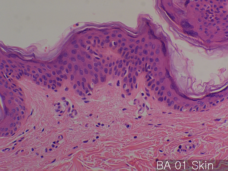


Evaluation slide (TEST SLIDE)

Product name Evaluation slide (TEST SLIDE)
Cat. No. BA
Current version BA4
Data sheet BA4.pdf
No. of samples 24
No. of patients 24
Core diameter 2.0 mm
Section thickness 4 micrometer
Description No. of normal cores: 12 cores
No. or cancer cores: 12 cores
The price of the TEST SLIDE is one-fourth of regular array slide.
The quality of this test slide is same as the other array slides.
This slide can be used for adjustment of experimental condition.
The TEST SLIDE is also useful for most researchers who consider
the experiment using tissue array slides.
Price 177 EUR
230 USD
150 GBP

Evaluation slide (TEST SLIDE) Skin Normal     Evaluation slide (TEST SLIDE) Breast NormalEvaluation slide (TEST SLIDE) Spleen Normal

Product Related Literature

A recent study of the tissue microarray, resulting in a rapid progress in toward the quantification of a large group of biomarkers in normal and diseased tissue. However, standard procedures for the collection of tissue has not yet been established for molecular profiling.

This study presents a high-throughput analysis of non-uniformity in the breast tissue of texture image for the purpose of identifying the region of interest in the organization for molecular profiling by tissue microarray technology. Image structure breast histological slides has been described with respect to three parameters: occupancy percentage ratio of the area occupied by image block chromatin (B) as the statistical heterogeneity index (P) and such areas, the stroma , in many cases, H is used for image analysis. It uses both the color and greyscale segmentation, is defined in the thousands per image block in the data set our texture parameters are calculated. The image block, were classified into three classes by using the parameters for the new texture features statistical learning algorithm. The categories are as follows. The image block these are not unique to a specific characteristic, to block cancer tissue, the disease state and normal breast tissue to normal image block.

Lead to the identification of tissue slides the same area as the cancer-specific grayscale and color segmentation technology. In addition to the image block that has been identified as a cancer-specific cells, and is part of the area crowded them in the section of the slide image of the whole was marked by two pathologists as the unit of interest for histological study further .

After 20 years, it is true has been proven, if available, the tissue Stanford Microarray Database today, images more than 200,000 are included, these advantages, clinical diagnosis related to tissue You write, and put a cloth colored with relevant organizations metadata. The TMAD, tools for the analysis and tools import score is included via a Web interface intuitive fabric design, and image.

Adaptive segmentation of histology images in constituencies they were applied in successive stages of the two. First, we used the index of the brightness to determine the (consisting of interstitial regions abundant chromatin) the foreground from the background. For this purpose, iterative algorithm is used to adapt the two component mixture Weibull parameters for monitoring the brightness. Algorithm iterative search online to find the mixing parameters that are optimized to fit the observed values. Then, the maximum likelihood threshold value between two components is determined by the brightness threshold value for separating the interstitial rich region is stained chromatin regions.

Several models of the system and object database that are disclosed, for managing the data organization. Purpose up to the modeling of metadata for comprehensive management of tissue microarray study of a large group. There are similarities, by providing public access to the raw data fabric experiment, TMAD is different. The ongoing collaboration with research group in the United States other than remove the sample slides during the delay and resulting complications and institutions have developed transported, a clear method for transferring metadata and images from cooperative organizations As a part.

Antibodies specific for the protein used for Human Protein Atlas project administered to tissue microarrays, to create a profile of expression and localization in 48 normal human tissues, based on the systematic creation of 20 types of cancer 47 cell line published Atlas of protein antibody-based public comprehensive.

Normal organs

Product name Normal organs in duplicates
Cat. No. AA
Current version AA9
Data sheet AA9.pdf
No. of samples 59
No. of patients 59
Core diameter 2.0 mm
Section thickness 4 micrometer
Price 244 EUR
320 USD
210 GBP

Normal organs      Normal organsNormal organs

Product Related Literature

Normal organs in duplicates can be evaluated by (PET-CT) scan and computed tomography (18F-FDG) positron emission tomography 18F-fluorodeoxyglucose in duplicate. I investigated the (women 32 men and 38 people) patients seventy people without the incorporation of non-physiological 18F-FDG. PET-CT before are within normal range blood sugar levels in the fasting state and image, all patients fasted for at least 4 hours. The image acquisition is performed after intravenous administration of 18 F-FDG, the image is obtained from the top of the thigh. Jeep various organs will be determined by the side view. Uptake of 18F-FDG is the highest in the spleen brain, cerebellum, heart muscle, tonsil, and liver in both men and women. There is information about the physiological uptake of Jeep authorities and normal 18F-FDG, but it is necessary for the correct interpretation of whole body 18F-FDG-PET-CT exam.

Using the colony assay in vitro clonogenic fibroblast cell precursors (CFU-F), are detected in the thymus bone marrow, spleen, and adult mice. CFU-F of total-body irradiation after D0 220, of 150 g reproduction of bone marrow, and is characterized by the delay time after irradiation and average loss, survival curves of CFU-F was irradiated immersion lasting from mouse bone marrow in vitro to return to the normal value to 25 days, the 6th is, following. This pattern of recovery after the transmission of the CFU-F is the same as the CFU-S. In addition, the number of CFU-F increased to about two times during the first 6 hours after irradiation.

Knowing the accumulation of FDG in the normal distribution for the interpretation of whole body PET imaging for the detection of tumor it is important. Review the whole body PET imaging, we studied the twenty-eight healthy people, which is a factor that led to the diversity and strength of FDG uptake in the major organs. Emission image is acquired, the image is reconstructed without attenuation correction. Were classified into four categories visually strength of FDG uptake. Accumulation, was not found thyroid, esophagus, the spleen within. I visualized the oral cavity, liver, stomach, large intestine, in all subjects. Muscles of the larynx of heart FDG card and neck muscles degree of 1-4 grade is different. Association, was not found between the intensity of the appointment of volunteer and age. The fasting, the heart, they were shorter in subjects who showed a “high” than shown (p <0.05) and significantly less accumulation. Significantly (p <0.05) also, the serum concentration of free fatty acids significantly lower these. Many organs, were observed in the heart muscle and laryngeal muscle, the neck especially the incorporation of a variety of FDG. In our study, there was no facter not cause FDG uptake in organs and the amount of free fatty acids present in the heart and the exception of fasting. This analysis of whole body FDG distribution in healthy subjects, is valuable for the detection of tumor FDG-PET.

It is a condition of congenital (oppositus location or horizontal stripes also called) Location inversion major internal organs or reversed, being reflected from the position of their usual. Normal mode is also known as solitus place. In a state that rarely other, known as heterotaxy doubt and place, it is not possible to determine the position.
Long-term position inversion is the short form of the phrase “where inversion viscerum” the Latin word meaning “reversal position of the internal organs”. After seeing first (heart is in the right side of the chest), and recognized by Marco Aurelio Severino in 1643, was painted by Leonardo da Vinci in 1452 to 1519 in the right chest. However, inversion which it was exercised, was described more than a century after the first by Matthew Bailey.
It varies between the populations that spread the location inversion is different, but it is less than 1 in 10 000.

It may be found in twins “mirror” the same or X-linked, but the inversion of the place, is an autosomal recessive genetic disorder in general. If congenital heart disease is not present, people are phenotypically normal in position inversion, it is possible to lead a healthy life without the usual complications associated with their medical conditions. Transpose place reverse hair loss in large vessels most often, there is a 5-10% the spread of congenital heart disease in patients. The incidence of congenital heart disease is at least 95% position of the inversion levocardia.

They know the abnormal anatomy of their people of many to seek medical attention for unrelated conditions, with inversion of hair loss whereabouts. Turning to the authorities, signs and symptoms of many “as a side ‘wrong then, can lead to some confusion. If you want to develop appendicitis, for example, people with a place reverse position, they to be a place to have their applications, will introduce the lower left abdominal pain doctor. Therefore, in the case of a medical problem, knowledge individual that is the position reverse position, be used to accelerate the diagnosis so that you can demand to shift to other signs and sounds of the heart. doctor can, people with this disease, wearing a. medical identification tags that can be used to notify the physician prior to testing You can tell the medical personnel in the event that the person is not able to communicate that you are