Ureter

Product name Ureter
Cat. No. 0073000A
No. of samples 1
Description ureter, normal
Age/Sex : 40/M
Price 197 EUR
260 USD
170 GBP

Ureter

Product Related Literature

In human anatomy, generate smooth muscle, ureter is a tube made of urine from the kidney to the bladder. In adults, long (10-12) 25-30 cm usually, ureter 3-4 millimeters – diameter. The histological, further smooth muscle layer of support peristalsis third and remote transitional epithelium is included in the ureter. In humans, before you descend to the bladder in front of the psoas major muscle, ureter, coming out of the renal pelvis inner side of each kidney. Ureter, across the edge of the pelvis near the junction (they are passing priority) and the iliac artery. This is a common place for the congestion (the rest of the ureter bladder transitional valve meets the pelvouteric junction and bladder meet the ureter renal pelvis, the renal hilum to ureter) of kidney stones.

Run to the wall of the bladder for a few centimeters, the junction of the ureter and bladder, ureter executes the side wall of posteroinferiorly pelvic curve anteriormedially then to enter the bladder in the back,. Return of urine is prevented by valves known as the ureter bladder transitional valve. I receive the uterine artery and mesometrium through the bladder ureter in women. “Water under the (vas deferens or uterine artery) bridge (ureter) the phrase” effective to remember this anatomical relationship is. different channels play the same role in fish and amphibians, but the ureter was found in species of amniote all other. You can move from the kidney, and kidney stones, is introduced into the urethra, this has the potential to cut off the flow of urine, causing the lower abdomen severe cramps, or back side,. It is blocked you can develop a hydronephrosis of the kidneys, kidney affected, has become swollen due to the flow of urine.
Usually, hysterectomy is a surgical removal of the uterus performed by the gynecologist. (Called removal of the uterus body, and, “supracervical” also, leaving the cervix intact), hysterectomy (removal of the body, cervical and bottom, often referred to as “full”) general or it may be partially. This is the Department of Obstetrics and Gynecology of surgery most commonly performed. 90% is made under the condition of benign hysterectomy more than 600,000, were performed in the U.S. alone in 2003. These rates are the highest in developed countries has led to considerable controversy hysterectomy is unfair, that has been carried out mainly because of the unnecessary. If you become a can not (as in the removal of ovaries and fallopian tubes) patients bear a child, is not available treatment options other you have the long-term effects and risks of surgery, removal of the uterus, the surgery is typically recommended. In many cases, there is a better option, it is, that the incidence of hysterectomy is reduced for non-malignant indications are to be expected.

In many cases, (removal of the ovaries) ovariectomy, is achieved using a hysterectomy in order to reduce the risk of ovarian cancer. However, recent studies that prophylactic oophorectomy without emergency medical instructions to reduce the long-term survival of women have severe side effects, substantially free of the other has been shown [4], this effect It is not intended to be limited to premenopausal women. However, women who have already entered have demonstrated that it reduced the long-term survival in ovariectomized after menopause.

Hysterectomy may be performed in various ways. Technology known the oldest, is the incision in the abdomen. Then, technology laparoscopic vaginal later (do a hysterectomy to vaginal canal) vagina (instrument inserted through a small opening near the navel often, additional) have been developed. This is done by (not to be confused abdominal incision, and laparoscopy) abdominal hysterectomy most in the United States. Usually, near the upper limit of the hair in the lower pelvis pubic individual like incisions made for cesarean but as possible, (Pfannenstiel) incision is carried through on the stomach wall side.

It enables greater access to doctor reproductive structures, this technology was carried out in order to remove the whole reproductive complex usually. Because of the need to cut through the abdominal wall by sometimes more, recovery time for a hysterectomy the open, is 4-6 weeks. Historically, the biggest problem with this approach was infections, infection rate, has become a major problem in modern medical practice of which is not well controlled. Open hysterectomy provides the most effective way for you to explore the abdominal cavity, to perform complex operations. Before analyzing the laparoscopic vaginal techniques vaginal and it also, in most cases, vaginal technique is the only possibility to achieve desired contrast subtotal hysterectomy.

With the development of laparoscopic technology of 1970-1980 period compared to the procedures of the abdomen, surgery and minimally invasive has gained great popularity among gynecologists “laparoscopic vaginal hysterectomy” is (LAVH) after recovery is much faster. In addition, it allows a slightly more complex operation and inspection good procedure of the vagina. LAVH is finished begin with laparoscopy, (with or without removal of the ovaries) as is done through the vaginal canal removal of the ultimate of the uterus. Thus, LAVH a total hysterectomy, you may need to be removed in the uterus neck. “Laparoscopic hysterectomy with the supracervical” (Rush) of the cervix using a divider that can affect the uterus into small pieces that can be removed from the abdominal cavity through the post-laparoscopic port without having to remove and has been developed for the removal of the uterus.

Kidney

Product name Kidney
Cat. No. 0071000C
No. of samples 1
Description kidney, normal
Age/Sex : 43/M
Price 197 EUR
260 USD
170 GBP

Kidney

Product Related Literature

Kidney is an organ that acts as an important regulatory some invertebrates including some in vertebrates most. They are essential in the urinary system, maintain the regulation of the electrolyte such that the acid-base equilibrium plays a homeostatic functions (such as vias to maintain the water balance and salt) regulation of blood pressure. Providing a body as a natural filter of the blood, they removes waste that is diverted to the bladder. The flow of urine, were separated, the waste, such as ammonia and urea, they also kidney is responsible for the reabsorption of amino acid water, and glucose. The kidney is, I produce the hormone, including enzyme renin calcitriol, and erythropoietin,. The discharged renal vein that receives blood from the renal arteries located in the back of the abdominal cavity to the retroperitoneal, kidney paired, paired. The pairing structure in the sky urine, the bladder itself to each kidney ureter.

While renal physiology is a medical professional relationship, kidney, is the study of renal function in kidney disease. Kidney is different, but often, people with kidney disease shows clinical characteristic symptoms. Clinical common symptoms, including kidney, including nephrotic syndrome and nephritis, renal cysts, acute kidney injury, chronic kidney disease, urinary tract infection, kidney stones, urinary tract obstruction. Various types of kidney cancer exists, kidney cancer most adult is renal cell carcinoma. It can be controlled by removing renal disease other tumors, cysts and, a nephrectomy or kidney. Measuring renal function may be always a bad case, dialysis, choice of kidney transplantation therapy by glomerular filtration rate as a. They are very harmful, kidney stones, can be annoying and painful. Then, removal of kidney stones involves sonication to divide the rock into smaller portions that pass through the urinary tract. Common symptoms of kidney stones one is acute pain in the inside next segment of the waist.

In humans, in particular channel lie peritoneum and paraspinal, I was placed into the abdominal cavity is located at a position kidney was slightly tilted angle. 2, there is one on either side of the backbone. Usually, the asymmetry is slightly lower than the left in the abdominal cavity caused by the liver leads to the right kidney, left kidney is disposed slightly inside the right. T12 spinal cord, the left kidney is about slightly lower-right level to L3. Just in the right kidney at the bottom of the rear and rear to the left liver and diaphragm, the diaphragm under the spleen. This is the adrenal gland is placed on top of each kidney. Part upper part of the kidney (the skull) are protected by 12 ribs and 11 partially, adrenal gland and kidney of the whole, is surrounded by (fat pararenal and adrenal glands) layer of fat in two, renal fascia each have. 155 g and between 115 men and between 125, each adult kidney weighs 170 grams in women. Left kidney is slightly larger than the right kidney is typically.

It has the structure of kidney bean shape, and having each kidney uneven surface. Concave, renal hilum is the point at which renal artery enters the renal vein or organ, left ureter. Fibrous tissue severe kidney itself is surrounded by fat adrenal perirenal fat, renal fascia and (of Gerota), are surrounded by the renal capsule. Between forward muscle horizontal stripes are the boundary line of the rear (rear) and (front) border of these tissues is the peritoneum. Edge is adjacent to the liver and spleen to the left kidney on the right kidney. Therefore, I will move down the intake of both.

In the renal cortex on the surface, is a deep brain kidney: kidney or real, material is divided into major two structures. Obviously, such a structure, each contained around the renal cortex of the brain called the (Malpighian) renal pyramids may be in the form of renal sheet cone 8 to 18. There are projections of cortex called (of Bertin) renal pyramids between renal column. Functional structure of the medulla and renal cortex cover nephron, and urine production. Pass tubule, initial filtering of the nephron is the renal corpuscle position, and cerebral cortex followed from medulla pyramid deep cortex. Some of the renal cortex, medulla rays, is a collection of the renal tubular drain to collect channel.

To empty the urine into a small cup, teat or tip, of each pyramid, minor calyx of the sky to the major calyces empty renal pelvis, is in major renal calyx and ureter. Renal artery while entering, is in the gate, renal vein and ureter, end the kidney. Is a lymphoid tissue and lymph node hilar fat about the structure of these. Is located next to the cavity filled with fat, called renal sinus fat is hilar. Calices and renal pelvis are included together renal sinus, I have separated these structures from renal medullary tissue.

The kidneys receive blood from the renal artery, the left and right from the abdominal aorta branches directly. In spite of the relatively small size of their kidneys receive approximately 20% of cardiac output. Branch of the renal artery, which penetrate the renal capsule leading to areas artery is divided further into interlobar arteries extend through the columns of the kidney during the pyramid kidney. Interlobar arteries after the blood flow to the arterial arch that passes through the boundary of the medulla and cortex. Each arc-shaped artery is supplying interlobular artery and some of its feed supply afferent arterioles the glomerulus. Interstitial space is functional in the kidneys filter the individual is rich in blood vessels (glomeruli). interstitum to absorb the liquid, which was recovered from the urine. In various conditions, may lead to the failure or renal insufficiency and scarring, which can lead to congestion in this area. Take place in order to move through a small network of veins filtration of blood was collected in between leaf veins after. to return to the leaf between veins come to form the end renal vein the kidney for transfusion as well as the arterial veins follow the same pattern of interlobular, to provide blood to the arcuate veins then.

Tonsil, normal

Product name Tonsil, normal
Cat. No. 0061100B
No. of samples 1
Description tonsil, normal
Age/Sex : 34/F
Price 197 EUR
260 USD
170 GBP

Tonsil, normal

Product Related Literature

Amygdala is a collection of lymphoid tissue. This term refers particularly to the palatine tonsil is a mass of lymph material located on both sides of the back of the throat of the man most commonly. Pharyngeal tonsils and tonsil of Palatine is a lymphoid tissue near the throat and pharynx. These fabrics, is a row of protective immunity first of the immune system against pathogens of inhalation or ingestion foreign. However, the role of the primary immunization of tonsils, it is to be understood yet.

Tonsillitis is a disease (pain and swelling) in tonsil inflammation. The most common way to treat it is an anti-inflammatory drug ibuprofen or when bacterial such, and antibiotics. Many patients dealing with tonsil removal of them at the tonsillectomy surgically. Collection of lymphoid tissue known as Waldeyer tonsillar ring including adenoid tonsil, two ectopic tonsils, two palatine tonsil, the tongue tonsils. The tonsils, tend to reach a maximum size of their adolescent close, they pass atrophy after gradually. However, they are the largest in terms of diameter of the throat in young children.

You can be, you can remove tonsils in the tonsils surgically and inflammation (adenotonsillar hyperplasia) and (tonsillitis) larger. If it interferes in patients with recurrent tonsillitis or frequently affect the absorption or the airway, can be observed this it. However, when tonsillar hypertrophy described, the mechanism of the pathogenesis of other subtypes of these two, it is possible to have different responses to the efforts of the same treatment. In elderly patients, may be an indicator of tumor or virus infection tonsils, such as lymphoma and squamous cell carcinoma asymmetric tonsils (also known as tonsillar hypertrophy of asymmetry), and the like. The hypertrophy of the tonsils, can be given the inability of sound velofaringealno and hypernasal, can affect the speech it. The size of the tonsils, can have a significant impact on upper airway obstruction in children with obesity compared to those of average weight.

One on each side, tonsils, is a small gland in the neck. They exist to help you fight bacteria when you’re a child. After the age of about three years, become less important in the fight against bacteria tonsils, usually reduced. Your body can fight bacteria without them still. They only take them if we are doing more harm than good. If we sore throat recurrence occurred despite treatment with antibiotics, they will take only tonsils. If they are large, to block the airway, the main reason for the other to remove the tonsils. Sometimes, small children, have a large tonsils so that they shut off the breathing at night. Parallel development in tonsil abscess, the result of infection of the tonsils, angina is the most unpleasant. People who have angina, choose to have a tonsillectomy to prevent another so often. If tonsils tumor is suspected, you are removed. If the increase tonsil tumor is expressed rapidly in size and bleeding ulcer and tonsils. Tumors of the tonsil is rare.

It is not necessary to remove the tonsils. You may want to check to see whether the it is only that the problem of the tonsils, you’d been waiting for. You need a doctor to explain why he or she feel that surgery is the best treatment he or she. By you can change your mind to work at any time, and to sign the consent form, it does not mean that there is a need to undergo surgery. If you would like, you can ask a specialist for a second opinion on the treatment. She will not bother to place it for you or him. You may have to arrange a second opinion to another specialist, you want to ask your own GP.

Tonsil surgery is very safe, all operations, has a small risk. The most serious problem is bleeding. This may require a second operation to stop it. This would have to be returned to the hospital of bleeding because there are two of Evert 100 children have their tonsils, and one of them will require a second operation. Such as, are you must tonsils of many of them is returned to the hospital because of bleeding, but only adults will require a second operation in the hundred. Of the 100 adults, during the operation of five, in particular, there is little chance that the tooth is chipped, there is a knock-out possible if we are crowned it or bukkake or are loose. You please let us know if you have any teeth of any such.

Pancreas, normal

Product name Pancreas, normal
Cat. No. 0059000B
No. of samples 1
Description pancreas, normal
Age/Sex : 64/M
Price 197 EUR
260 USD
170 GBP

Pancreas, normal

Product Related Literature

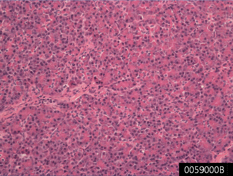
The pancreas is a gland organ in the endocrine system and digestion of vertebrate. Generates important hormones, including some pancreatic polypeptide insulin, glucagon, and somatostatin secretion pancreatic juice containing digestive enzymes that promote digestion and nutrient absorption in the small intestine, endocrine glands, is also in the digestive organs it. These enzymes help to break down carbohydrates, proteins and lipids in the chyme further. Under the microscope, the sections of the pancreas were stained, indicating different types parenchyma, two. Slight staining groups of the cells is referred to as producing islets of Langerhans hormones that defines the endocrine function of the pancreas. Darker staining cells, to form a bunch that is connected to the tube. Belongs to the intestinal digestive enzymes through a system of channels, acinar cells of the exocrine pancreas secretes.

Provides the exocrine and endocrine pancreas is a dual function gland. Portion of the pancreatic endocrine function is approximately 1 million cell clusters called islets of Langerhans. Main types of single cell 4 is present in the islet. Be distinguished using staining techniques standard is relatively difficult them, be classified by secretion of their can they: (elevated blood glucose) glucagon secreting cell α, the β cell gamma cells and PP cells in the pancreas that delta cells secreted polypeptide secretion (decrease in blood glucose levels) insulin secreting, somatostatin (and control / stop α β cells) and.

Is a collection compact endocrine cells arranged in clusters and code, it is twirling in dense networks capillaries island. The capillaries of the islets, and backed by a layer of endocrine cells in direct contact with the container, through the cytoplasm or process, are in direct contact with blood or endocrine most cells is due to direct printing. By Alan E. nurse, depending on the volume of the body, and their production of “hard hormone island is usually around pancreatic cells of them as if they were placed in the part of a completely different part of the body islets is “ignored, plays an important role in glucose metabolism, and regulation of blood glucose concentration. I will help the digestive system as exocrine pancreas. It secretes pancreatic fluid containing digestive enzymes to pass through the small intestine. These enzymes help to break down (fat) lipid carbohydrate, and protein in the chyme further. In humans, direct, secretory activity of the pancreas is regulated by the action of the autonomic nervous system in blood flow indirectly by the action of the hormone in the blood of the islets of Langerhans.

Secretion of digestive enzymes such as amylase and lipase, such as the abdominal cavity, the potential and lead to damage to the organs inside the abdomen and pancreas digestion and self-digestion of then, usually, excavation of the pancreas, medical intervention experienced rapid and I need. Inflammation pancreatitis, the pancreas. Various factors cause the high pressure of the pancreatic duct. Pancreatic duct leakage and rupture of pancreatic juice causes the self-digestion of the pancreas. Therefore, pancreatitis occurs. Gallstones and alcohol is the most common cause of two of pancreatitis. Pancreatic cancer, pancreatic cancer, exocrine, mortality is very high to be one of the worst cancer in particular. Pancreatic endocrine tumor is rare. Representative: insulinoma gastrinoma (5% 95% benign, malignant).

It is a chronic autoimmune disease (also known as juvenile diabetes), immune system type 1 diabetes attacks the insulin-secreting cells of the pancreas. This results in a blood sugar level of patients rise to a dangerously high level. To fix this, the patient should take the third day insulin shot. In addition, there may be a correlation between some between pancreatic cancer and diabetes, and chronic pancreatitis. Type 2 diabetes is more common in adults overweight, but are seen in children too. Unlike type 1, it can be adjusted by the weight-loss drug continuously it. It may be if they are administered insulin to regulate the concentration of pancreatic enzyme supplements for patients aid digestion and blood sugar properly without pancreas is alive.

Gallbladder, normal

Product name Gallbladder, normal
Cat. No. 0057000A
No. of samples 1
Description gallbladder, normal
Age/Sex : 52/M
Price 197 EUR
260 USD
170 GBP

Gallbladder, normal

Product Related Literature

In vertebrates, the center helps to digest fat (gall bladder, gallbladder, bile duct vesicles) The gallbladder is a small organ concentrate bile production in the liver. In humans, in most cases, loss of the gallbladder, may be easily tolerated. Surgical removal of the gallbladder, is called cholecystectomy. Gallbladder is a hollow system located beneath the liver exactly. In adults, the gallbladder, measuring about 8 cm and length (3.1 inches) diameter 4 centimeters (1.6 inches) when it is fully deployed. Bottom neck, body: it is divided into three parts. Connect to the bile duct through the binding of the cystic duct hepatic duct common to become the common bile duct and then taper neck. Gallstone is called a stuck bag Hartmann often neck of the gallbladder mucosa times. Angle of the gallbladder is disposed between the lateral edge of the rectus abdominis muscle and coastal margin.

Gallbladder disease, is a disease related to the gallbladder. You may develop in other parts of the same system in the bile gallbladder gallstones. If can not gallstones gallbladder is symptomatic, it is divided into smaller pieces by the ultrasonic dissolution or with the drug, surgical removal of the gallbladder, known as cholecystectomy may be mentioned. Degradation products of gallstone bacteria of biliary lipid thin sheet of solubility of biliary lipid board, or unconjugated bilirubin, occurs in favor of precipitation of cholesterol when. For cholesterol gallstone cholesterol secretion of liver, metabolic diseases, intestinal bacterial degradation of bile salts in order to destabilize the cholesterol carrier in the bile, to produce crystals of cholesterol in combination with the change in motion of the gallbladder . The case of black pigment gallstones, changes in absorption and metabolism of heme lead bilirubin, increased bilirubin concentration and deposition of bilirubin calcium. Is the main factor leading to precipitation and bacterial degradation of brown pigment stone biliary lipid as opposed to mechanical obstruction of the biliary tract.

In a particularly fertile year, women, is an organization with the most compelling gallstones. Men to form a stone, may almost double, start for people to catch up, the difference narrows after menopause women. Female hormones, parity, estrogen replacement therapy or birth control pills, the main mechanism is the well-established risk factors for cholesterol gallstones. Female hormones, an adverse effect on the secretion of the liver function of the gallbladder and bile. Increased the secretion of cholesterol, reduce the secretion of bile salts, due to damage to the blank, bile salts, and estrogen reduces the secretion of progestin action gallbladder leading to shutdown. Progestin mud speaker Lennon of the new fourth generation to be used in oral contraceptives some increase the risk of cholecystectomy and gallstones further, but a fairly modest in the increased risk, the possibility that can not be clinically-important There is sex. State of Michigan that is compared with the 341 478 non-users, 138 943 user retrospective (historical) cohort study of the OC line in a large database that contains the data for 1981 and 1980 for billing Medicaid in Minnesota Broken. And high enough to be a problem clinically in young women only risk, but birth control pills, that it is a risk factor for gallbladder disease has been shown.

Those predisposed to it, some acceleration of the disease of women, but in 1984 the Royal College of General Practitioners oral contraceptive research, long-term oral contraceptives, shows that it is associated with an increased risk of gallbladder disease. In a recent study, it is suggested that this is not the case. The 1993 meta-analysis, it was concluded that the use of oral contraceptives, and is related to a transient increase incidence and mild gallbladder disease, good buying and selling, the low-dose oral modern effect even contraceptives, check exclusion is safer than the old formulation. [8]
In 2001, in the comparative study of IMS Health lifelink planning, risk of gallbladder disease associated with norethindrone comparison desogestrel, and drospirenone interpretation database as a large group of women using oral contraceptives is small, It was claimed that levonorgestre statistically significant increase can not be found. There is no statistically significant increase in risk associated with (ethynodiol diacetate, norgestimate and norgestrel) Other formulations of oral contraceptives.

There is a possibility that (1cm or less) is not a cancer Ost small polyps, unchanged for years. However, small polyps, for example, when viewed in other conditions such as primary sclerosing cholangitis, they likely to be benign low. Likely to develop into cancer, the higher the large polyp. Cholesterolosis is characterized by the growth of the lining of the gallbladder in the prediction of fingers due to excessive accumulation of cholesterol and triglycerides in intraepithelial macrophages. Cholesterol these polyps are benign gallbladder polyp most. The adenomyomatosis, other too thick, gallbladder wall discloses a pathological state of the gallbladder by the distribution of ground cell layer. This is characterized by a deep fold in the proper muscle layer. Called Rokitanski-Aschoff sinuses, ultrasound examination may reveal a campus diverticulae thickened gallbladder wall.

Liver, normal

Product name Liver, normal
Cat. No. 0056000C
No. of samples 1
Description liver, normal
Age/Sex : 75/F
Price 197 EUR
260 USD
170 GBP

Liver, normal

Product Related Literature

The liver, an important organ in animals and some other vertebrates are present. It has a wide range of functions, including detoxification, protein synthesis, and production of bio-chemical and necessary for digestion. It can be used in the short term new technologies liver dialysis, a method of compensating required for survival, the lack of long-term liver function, there is currently no liver. It plays an important role in metabolism, glycogen storage, disintegration of red blood cells, plasma protein synthesis, hormone production, and the body has a number of functions in the body, including detoxification. It is located below the diaphragm in the abdominal-pelvic region of the abdomen. It produces alkaline compound to help digestion by emulsifying bile, the lipid. Many of them, small is necessary for the normal function of life, organization highly specialized liver is to regulate a wide range of mass biochemical reactions such as decomposition and synthesis of complex molecules.

The liver is a reddish brown organ with four leaf shape and unequal size. Human liver, has soft weight and 1.44 to 1.66 kg of normal (3.2 to 3.7 kg), brown tinged with pink, the organ of the triangle. (Skin is the largest organ overall) and the internal organs of the maximum, this is the largest gland in the human body. It just resting on the diaphragm below, I am located in the upper right quadrant of the abdominal cavity. Liver is located to overlap gallbladder and right stomach. Is connected to the large vessels of the two, it is called what’s called portal vein and hepatic artery one. Not only the entire intestinal tract, whereas carry blood containing the nutrients absorbed from the pancreas and spleen, hepatic artery carries blood from the aorta to the portal vein. These vessels subdivided into capillaries lead to small leaves then. Each lobule is made up liver cells, which is the main metabolic cages, with several million people. Unit is the functional unit of the liver.

Long-term bile duct was prepared from the woody branches biliary. Is collected in the merge bile canaliculi to form a bile duct bile produced by the liver. In the liver, ducts are called (in the liver) bile duct liver, they are considered to be (outside the liver) liver once, and then exit the liver. The merged to form the hepatic duct common Finally, intrahepatic duct discharging to the left and right hepatic duct. Cystic duct from the gallbladder joins the liver of common duct to form the common bile duct. The duodenum and through stored in gallbladder temporarily through the cystic duct or bile duct or bile duct directly. Pancreatic duct and common bile duct enter the second part of the duodenum in the ampoule along the Vater.

When viewed from the gross anatomical top surface of the additional splitting the liver (left and right) of two parts, but if there, is divided into four parts visceral surface plus the tail lobes square. A link that curved looks (front side) front of the liver. This separates the anatomical right leaf anatomy and the left liver lobe. If you turned to look at it from the liver (visceral surface) below, there is a partition of two additional between the left and right. Tail lobe and (high) square leaf These are (bottom).

Determine visceral surface functional anatomy, how to organize the liver. Imaginary line that runs the gallbladder split in half and all the way to the inferior vena cava from the left you should see one. This line is called the line-scan her. This line divides the liver to the left and right. (Ligament of Aranshio) venous ligament such as round ligament (ligament teres), anatomical and other characteristics, are still separated on the left side of the liver sections of two more. Now, an important anatomical landmark, horizontal slit of the liver, separates this part of the left segment of the four numbers starting from the tail lobule as well as the anti-clock mode is attached, of the liver. Eight segment will only be seen top view, from this point of view, we can see the 7 segment of the internal organs. It is composed of units of each of the lobes, is then run from binding around the hepatic veins to carry the blood from the liver vein.


Parotid gland, normal

Product name Parotid gland, normal
Cat. No. 0055100A
No. of samples 1
Description parotid gland, normal
Age/Sex : 37/F
Price 197 EUR
260 USD
170 GBP

Parotid gland, normal

Product Related Literature

Parotid salivary gland is a human. A double-stranded structure, which is the maximum of the salivary glands. In order to facilitate swallowing and chewing and in order to initiate the absorption of starch, which is wound around the saliva and separated by a channel ramus Stensen in the oral cavity. The word “mumps” means around the ear (paraotic) literally. Under, parotid gland is a pair of serous salivary glands located in front of the ear canal to drain the secretions of their own in the vestibule of the mouth through the tube Stensen her. They are the major salivary glands of the largest of the human body.

Nflammation of one or both of the parotid gland is also known as the mumps. The most common cause of parotitis is mumps. A wide range of vaccination against mumps has reduced the incidence of measles mumps significantly. Regardless of external pressure and salivary duct stones from other infections viral infections, such as bacterial infections such separately, can cause (mumps, chronic suppurative parotiti or acute) the mumps as the cut-off channel . There is a possibility that syphilis and tuberculosis also cause granulomas formtation parotid gland. Main confluence of the main duct and stone occur in the main parotid duct. The saliva, when there is a tendency to avoid foods that produce this condition, patients usually complain of pain. In addition, you may want to become enlarged as trying to eat the parotid gland. Pain can be reproduced in the clinic squirting lemon juice in the mouth.

In surgery is a section of the oral mucosa and normal sphinterotomy to permit removal, the front of the channel depends to if the location of the stone, but if it is disposed on the back of the main duct, it must be completely resected glands There is a case to be. May be due to lymph benign lesions caused by Sjogren’s syndrome and Mikulicz disease is swelling of the parotid gland. Create and swelling of the parotid gland, the appearance of the line of heavy jaw, it indicates a failure bulimia nervosa, you may want to eat. 80% of eighty-tumor of the parotid gland are benign. The most common of these is (ie gland lymphoma) Warthin tumor and pleomorphic adenoma of many men than women. Its importance is related anatomically, it is a tendency to increase over time. The growth of the tumor, there is a potential to change the order of the gland, facial nerve causes facial pain on the side of traveling through the gland.

Mucoepidermoid cancer and tumor, 20% from 15 minutes of parotid gland tumor is adenoid cystic carcinoma of the most common malignant. Cancer of undifferentiated carcinoma parotid pleomorphic adenoma chemical cell carcinoma, cancer of EX, (pancreatic duct epithelial origin of parotid) glands, and, (resulting from real parotid gland) squamous cell carcinoma includes other cancers are. Critically, must be defined as a resection of the tumor is related to a branch of the facial nerve (CN VII), in the paralysis of the facial muscles of the face, as a result, there is a possibility of damage to the nerve. I increase the likelihood of recurrence after surgery and in some cases difficult anatomical relationship of the facial nerve parotid gland rental because of the surgical treatment of tumors of the parotid gland. Therefore, detection of early stages of parotid gland tumor is very important from the point of view of prognosis after surgery. Incomplete before treatment made in a range of other professional and cause attacks surgery technology is difficult.

Innervation is completely autonomous. Reached around gland periarterial plexus of the external carotid artery postganglionic sympathetic fibers from the superior cervical sympathetic ganglion, its main function is vasoconstriction. The cell bodies of preganglionic sympathetics, usually in the corner next to the spinal cord segment of the chest at the top. Sympathetic fibers in the glossopharyngeal nerve section before, I leave the brain stem of small salivary nucleus eardrum, through the next lower cone branch path to the otic ganglion then. They synapse to reach the gland hitchhiking through branch auriculotemporal nerve, mandibular nerve postganglionic fibers there.

Lung, normal

Product name Lung, normal
Cat. No. 0028000B
No. of samples 1
Description lung, normal
Age/Sex : 62/M
Price 197 EUR
260 USD
170 GBP

Lung, normal

Product Related Literature

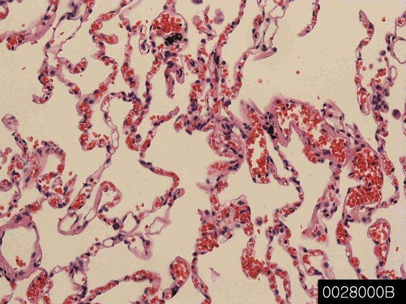
It is the breath of the most important body in the air-breathing animals, including many four-legged most, a snail and a few fish in a small number of lung. Mammals, in a more complex form of life, close to the spine on both sides of both lungs, the heart. Its main feature is that in order to transport oxygen from the atmosphere into the bloodstream, releasing carbon dioxide into the atmosphere from the blood. This exchange of gas will require a large area to be achieved little by the mosaic of specialized cells that form millions of thin-walled air sacs very called alveoli.

In order to understand the anatomy of the lungs, to consider the air passage from the nose and mouth of the alveoli. Passage of air through the nose or, mouth and pharynx through the nose, pharynx, larynx, trachea (windpipe). In the system of bronchial they reach the bronchoalveolar lavage gradually, fragmented, air passes through the trachea split into two branches bronchi of these lungs. It is alveoli these many places gas exchange oxygen and carbon dioxide is performed. In tetrapod breathing fast, is driven by muscle activity, it is still, the air was driven high by the muscles of the pharynx through the pump in cheek amphibians. Reptiles, birds, mammals uses the musculoskeletal system in order to support breathing, and promote. Or pneumatic or Latin pulmonarius,: in the medical terms related to the lung, starting (high adjective form), in the lungs frequently.

The lungs of mammals, including humans, has a soft texture and sponge-like, the number of the outer surface of the lung itself is a large area much, you have the epithelium of honeycomb. Largely due to the diaphragm muscle at the bottom of the breast is large breathing. Guide the airflow in the airways, by volume, therefore, contraction of the diaphragm pulls the bottom of the cavity which is closed to the lungs in order to increase the pressure drop it. The air enters through the nasal cavity and oral cavity, then, in any branch within the department and subsequent main bronchus, it goes through the pharynx, the larynx and trachea. During normal breathing, expiration is a passive, muscle is not contracted (the diaphragm relaxation) and at all. Rib cage itself can be expanded to a certain extent by the action of the muscles of the respiratory tract and other accessories and contract. As a result, the air expelled from the lungs, or be transported inside. This type of lung similar to the bellows of a blacksmith, known as bellows lung it.

In humans, I was divided into two bronchi that enter the root trachea and lung. It continues to divide bronchi and lungs, leading to bronchial after several departments. It will continue the bronchial tree branches until it reaches the level of terminal bronchioles leading to the alveolar sacs. Alveolar sacs called plural form of infundibulum is made up of a group of alveoli like grape individual within the bundle sometimes. Alveolar individual is wrapped in blood vessels strictly is here that gas exchange actually occurs. Deoxygenation oxygen is pumped through the pulmonary artery to the lungs diffuse into the blood and blood from the heart and replaced with carbon dioxide in hemoglobin in red blood cells. Oxygen-rich blood returns to the heart through the pulmonary veins to be pumped back into the systemic circulation. “Health center and respiratory lung disease”.

And is disposed in a chamber of the two in human lung, on both sides of the heart. Appearance is similar, but the two are not the same. Both of the two left and right lobe of 3, it is divided into units of crack. Then a segment unit is a minimum unit visible to the naked eye, the sheet is divided into sectors hexagonal lungs. Black and connective tissue separating the unit in smokers frequently. Inner edge of the right lung is substantially vertical notches of the heart while include left lung. In order to correspond to the shape of the heart, the notch of the heart, is a concave impression molded. Each title will be surrounded by a pleural cavity, pleural consists of two. Parietal pleura is located in the chest, visceral pleura is located on the surface of the lung. In between the pleura is pleural effusion.

Pleural cavity helps it to lubricate the lungs, is maintained on the surface of the lung in contact with the chest to provide a surface tension. Somewhat lung is “exploitation”, it has a huge reserve capacity as compared to the exchange of oxygen at rest. While moving slowly or still, even if there is no significant reduction in lung function in such a situation, excess capacity such, is one of the reasons why we can U smoke for years people it is, only a small portion of the lung, and blood gas exchange, it has not been flooded actually. Destruction of alveolar many situations lead linked to extreme shortness of breath, emphysema, too over time. By oxygen requirement to exercise the lung volume big, to be able to make the body fit the requirements CO2/O2 course if you increase, you are submerged. People with offset each other at a loss, further, as a result of capacity, it is possible to live in the lung only.

Bronchus, normal

Product name Bronchus, normal
Cat. No. 0026000A
No. of samples 1
Description bronchus, normal
Age/Sex : 37/F
Price 197 EUR
260 USD
170 GBP

Bronchus, normal

Product Related Literature

Bronchus is a passage of airway in the respiratory tract to go to the air (multicast bronchial bronchial adjective) lung. In bronchial branch, a small tube made in order to bronchi. No gas exchange does not take place in the first part of the lung. Anatomy Depending on the mode human trachea (windpipe), known as Carina is divided bronchus of the left and right two (also mainstream bronchi) and, in the two vertebrae level or second pentagonal thoracic sternum and higher or lower breathing in the histological point. Right main bronchus is wider than the vertical left main bronchus, more, shorter. The right main bronchus, it is subdivided into lobes bronchi of three, the left main bronchus, and is divided into two.

Known segmentalinic bronchi, as each supply bronchial segment in the lobar bronchi, were separated in tertiary bronchus on. It is a division of lung bronchial segment is separated from the rest of the lung from the barrier of connective tissue. This feature allows the bronchial segment to be deleted without affecting the other segments surgically. Segments of the lungs of 10 leading to 8, due to the anatomical development, there are some segments of the bronchi of the left lung fuse. Composite bronchus extends to divide the 2-11 lung ducts then generates a respiratory bronchioles, some split the terminal bronchioles in each of which, to divide the main bronchi plurality. There are five, and six alveolar sacs associated with each alveolar duct. Alveoli is the basic unit of anatomy pulmonary gas exchange.

Giving a shape like a small island in the small bronchi and plate and the large bronchi in the form of “D” to them, hyaline cartilage to form an incomplete ring in the bronchi. Smooth muscle is present continuously in the peribronchial. The level mediastinum, the fourth thoracic vertebra, the trachea, were divided into right and left primary bronchi. Smaller branches of the bronchi in the channel by the end of the small air sacs called alveoli. Main bronchial mucosa and cartilage are the same as those of the trachea. Bronchial tree; therefore followed by a branch, the amount of hyaline cartilage is reduced over as bronchial wall. Cartilage decreases as the amount of smooth muscle increases. Lining undergo the transition to a simple squamous epithelium to a simple cubic epithelium from eyelashes pseudostratified columnar epithelium.

Simple squamous epithelium that allows for fast diffusion oxygen and carbon dioxide in the alveoli and alveolar tube, and consists essentially. Gas exchange between the air in the blood and lung capillary is held in place by the wall of the alveolar ducts and alveoli. Bronchitis, which is defined as inflammation of the bronchial tubes. There are two main types of chronic and acute. Usually, acute bronchitis is caused by a bacterial infection or virus. Chronic bronchitis is a form of chronic obstructive pulmonary disease, which is usually associated with prolonged exposure to a stimulus and smoking. In many cases, asthma is bronchial hypersensitivity having an inflammatory component in response to allergen.

Skeletal muscle, normal

Product name Skeletal muscle, normal
Cat. No. 0013000A
No. of samples 1
Description skeletal muscle, normal
Age/Sex : 61/M
Price 197 EUR
260 USD
170 GBP

Spleen, normal

Product Related Literature

Striped muscle tissue under the control of the somatic nervous system, i.e., skeletal muscle, is a form of voluntary control. This is one of the others three major types of muscle, and smooth muscle and myocardium. As its name suggests, is attached to the bone of the package and is composed of collagen fibers, known as tendons.Skeletal streaks individual elements known as “muscle cells” or muscle cells of skeletal muscle, called “muscle fiber” colloquially sometimes. . In the process known as myogenesis, they are formed (lead-type embryonic stem cells, muscle cells) from the fusion of the development of myoblasts. These long, cylindrical, multinucleated cells called muscle fibers.

Muscle fibers, are made up of muscle fibrils in order. It consists of myosin filaments and actin repeat sarcomere of myofibrils, as the basic functional unit of muscle fibers, responsible for the appearance of skeletal muscle strip, which provides basic facilities needed for muscle contraction. Means multiple packets of integrated muscle fibers by the terms muscle connective tissue. Muscle individual fibers are long, and is formed during the development of fusion undifferentiated immature cells of several known as myoblasts cylindrical into polynuclear cells. Discrimination in this state to complete mainly before delivery of cells, the size of the then continues to increase. When viewed under a microscope for a position in the cytoplasm cytoskeletal component of muscle fibers, skeletal muscle showed stripes unique pattern. It is the major cytoplasmic proteins (known respectively “thin filaments” “as a” thick) actin and myosin are arranged in repeating units called sarcomeres.

Interaction of actin and myosin is responsible for the contraction of muscle.
Degree of oxidative phosphorylation (fast or slow) the type of myosin is present, fiber passes: You can sort the muscle fibers, there are two basic ways. The skeletal muscle, be divided into two categories in this way you can: Enter the type II with me. Enter the I fiber is displayed in red due to the presence of oxygen-binding protein myoglobin due. Because use oxidative metabolism to generate ATP, they are suitable for slow fatigue resistance and fibers. Because myoglobin is not present, it is white, type II fibers is dependent on the glycolytic enzyme. For such fibers are effective for short bursts of speed and power, is used as the anaerobic metabolism of a specific subtype with the oxidative metabolism. There is a fatigue quickly these fibers.

Red fibers: those containing a higher level of oxygen storage protein myoglobin was the red appearance. The red muscle fibers, tend to have a capillary density and higher local mitochondria. White Fibers: Low of these were white appearance. As to learn about the functional differences between the skeletal muscle fibers, characteristic spasm of slow muscle at high speed, are classified according to the means of them, most, not overlap the previous classification completely, colors or more Based on the. Fast muscle: Some authors, will be able to split the ATP very quickly definition of fast muscle fiber to that of myosin.

However, also fast muscle fibers, and provides greater capacity than sending the electrochemical potential of the release rate of calcium and fast action, it is taken from the sarcoplasmic reticulum. Fast muscle fiber can be used to reduce the voltage of 2-3 times higher than the proportion of slow twitch fiber, and grow well developed for the transmission of energy, and are dependent on glycolysis short-term . By generating the short bursts of speed and strength than slow muscle, it is much better so fast muscle fatigue of fast muscle.

Slow twitch: slow twitch fiber to generate the re-synthesis of energy for ATP by long-term system of aerobic energy transfer. They tend to have a low level of activity of the proton pump at a slower rate contraction with a better solution sugar development capability. Give the red pigmentation to them, and a high level of mitochondrial number and large myoglobin is included in them. They, thus to have they are fatigue resistant, high concentration of mitochondrial enzymes has been demonstrated. You can be taken long before muscle fatigue of slow muscle is firing from fast muscle fiber more slowly.

The two main categories of muscle fibers, based on the myosin isoforms, there are several differentiation of type II in several subtypes are further denoted by a letter of the alphabet. In humans, there Pa then two subtypes is in IIx of, the IIx, often referred to as the classification of the previous IIb and are clustered in two different types. Then, further research, these subtypes are recognized as different, but using the first name IIb, remained to fixing. Fiber type of non-human, are included IIb fibers of the actual, the IIc, and IID,.

Macromolecules muscle fibers, are arranged so as to verify that it meets the form and functions organelles. Plasma membrane is called the cytoplasm muscle sheath known as the sarcoplasmic. There is a muscle fibrils in the sarcoplasmic. Long link protein of about 1 micrometer in diameter of muscle fibrils, is the muscle filaments, each containing. Pressed against the inside of the muscle cell membrane has to flatten the nucleus abnormally. There are mitochondria in muscle fibrils between. Muscle fibers is not a smooth endoplasmic reticulum, but it contains the sarcoplasmic reticulum. It holds a reserve of calcium ions required for muscle fibrils of sarcoplasmic reticulum surrounds, to induce muscle contraction. On a regular basis, will be expanded, the bag end known as a terminal Shisutane it. They are, across the muscle fibers from one to the other. Shisutane between the two terminals, is the mass transport of cylindrical called horizontal tubular and (T tubules). T tubules is the possibility for action for notifying the sarcoplasmic reticulum to release calcium cause muscle contraction. Together, side tubules and terminal Shisutane the two form a triad.