Kidney renal cell carcinoma clear cell type

Product name Kidney renal cell carcinoma
Cat. No. 7183123A
No. of samples 1
Description kidney, renal cell carcinoma
Age/Sex : 62/F
Price 197 EUR
260 USD
170 GBP

Kidney renal cell carcinoma clear cell type

Product Related Literature

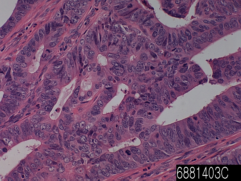
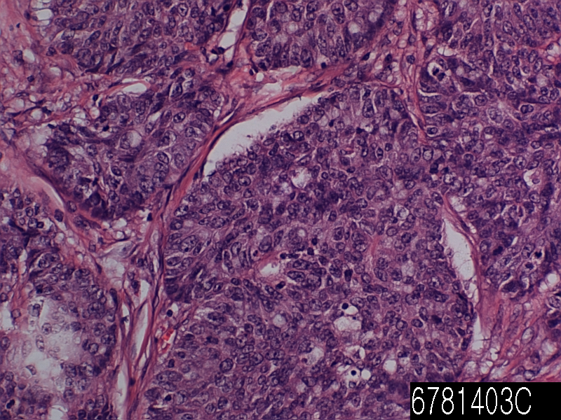
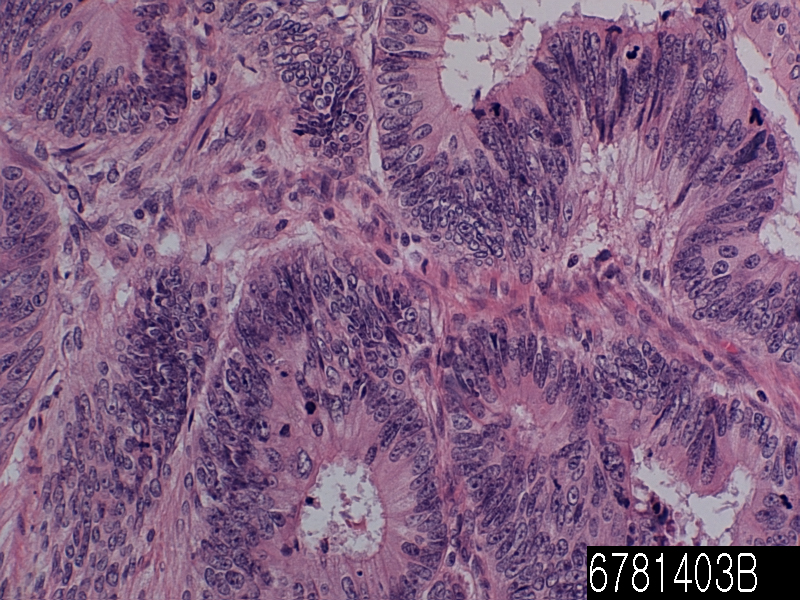
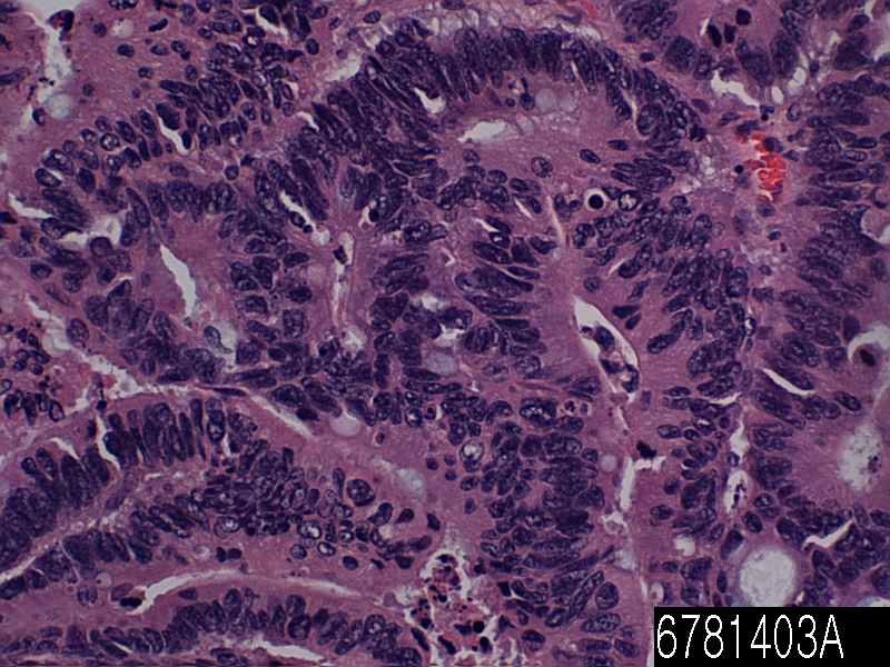
Clear cell renal papillary carcinoma, cancer of the kidney known as clear cell tubular papillary renal cell carcinoma and even abbreviated the CCPRCC, have a microscopic morphological features papillary renal cell carcinoma, clear cell kidney cancer It is the type that has been described recently, immunohistochemistry and molecular changes is important pathologically based on it. In other words, the core to have a nuclear cusp is in close proximity to the aspect of the lumen CCPRCC classic. typical structure of the gland most basally located nucleus, that is, the cytoplasm in the immediate vicinity of the basement membrane.

(Known as adrenal also RCC,) renal cell carcinoma, it is derived from kidney cancer in the lining of the renal tubules of the nearest, to kidney very transport of GF glomeruli descending part of the nephron small tube (glomerular filtration). Accounted for about 80% of cases, RCC is the most common type of kidney cancer in adults. This is described as one of the most lethal of cancers urological all. Is a partial nephrectomy or radical most commonly, initial treatment remains of the pillars of health care. Tumor is limited to the renal parenchyma, the 5-year survival rate is 60-70%, but this was significantly reduced, wherein the transition is spreading. This is the response of some cases of immunotherapy, but there are relatively resistant to chemotherapy and radiation. Targeted therapy sunitinib, temsirolimus, bevacizumab, and sorafenib and interferon-α, such as this has improved the outlook for RCC.

Daniel Zeneru Prefecture, the first reference, suggested a tumor that occurs in the kidney in the text Practicae Medicinae, which was announced in 1613 first. Miril has announced a clear case of early renal cell carcinoma in 1810. In April 1809, he suggests that it is in the later stages of pregnancy, and describes the case presentable Francoise quiet to City Hospital of Brest on the 6th, the 35-year-old woman. Koenig has announced the initial classification of renal tumors based on the macroscopic form in 1826. Koenig Split medulla form scirrhous tumor, steatomatous, and fungi.

After classification of tumors, researchers have attempted to identify the tissue of origin of the renal cell carcinoma. Pathogenesis of renal epithelial tumors, one of contradiction most enduring of surgical pathology modern provided. Discussion in 1883, he was started by Paul announced Gras Witz, small, his observations of morphological yellow kidney tumor. Grawitz has entered into a lung tumor only those of adrenal origin papillary tumor, whereas renal tissue of. 1893, Poruzudekku atrophy, published a description of the renal tumor have mentioned some atypical features prominent gradient of atypical features of these between the malignant tumor that he and the adjacent tubule and tubule I challenge the assumption that theory by Grawitz with. Support in 1894 Otto Lubarsch,, assumptions theory coined by Grawitz long-term hypernephroid tumor, which was revised in adrenal of Felix Victor Birch-Hirschfeld to describe these tumors. It is considered the origin of the adrenal gland of renal tumor, sharp criticism Grawitz offer has not been proven in Stoerk Oscar in 1908. In spite of the argument convincing to assume that theory by Grawitz, adrenal related connotation and long-term adrenal continued in the literature.

Such as foot and Humphries, and foot. And coined the term single cell cancer renal stress in renal tubular origin of these tumors. Their names will be changed to long-term renal cell carcinoma widely accepted already Fetter little. Conclusive evidence to resolve any dispute has been proposed by Oberling et al. 1959, people who have studied the fine structure of clear cell renal cell carcinoma 8. The cytoplasm of tumor cells, they found that deposits and mitochondria contain a lot of fat and glycogen. They identified the cell membrane which is arranged perpendicular to the membrane cells and occasional containing microvilli in free coverage. They are characteristic of these tumors was concluded that indicate that arise from epithelial cells settling and renal tubular any of the issues discussed in tumor pathology finally determined by it.

Historically, doctors expect the person to submit what is known as a classic triad of symptoms. 1 hematuria is when there is blood in the urine contains: this triad of symptoms. 2 abdominal pain is a pain in the side of the body between the ribs and femur. It is similar to three abdominal mass, and fullness, but big. That this triad classic symptoms of current only occurs in 10-15% cases are known, which may indicate renal cell carcinoma (RCC) in the advanced stages. Today, (mostly means that there is no symptom) is a fairly asymptomatic, people are detected in a test accident for other diseases usually RCC.

Rectum adenocarcinoma PD

Product name Rectum adenocarcinoma PD
Cat. No. 6881403C
No. of samples 1
Description rectum adenocarcinoma PD
Age/Sex : 55/M
Price 197 EUR
260 USD
170 GBP

Rectum adenocarcinoma PD

Product Related Literature

Seen in young women in general, and have a better, VGA high differentiated adenocarcinoma villoglandular villoglandular papillary adenocarcinoma, and papillary villoglandular adenocarcinoma, has been shortened, as well as other cervical adenocarcinoma Villoglandular, in relation to cervical cancer, prognosis is a rare type of cervical cancer. Exhibition compression replaced papillary adenocarcinoma, tumor, normal lung tissue and form papillary structure complex, is the organizational form of lung cancer that is malignant cell growth diagnosis of destructive. The name of the lesions, describes the microscopic appearance of it. It is covered with glandular pseudostratified columnar epithelium, it has a structure like nipple and (papilla) long fibrovascular core and its relation to the width, (such as villi). The earwax adenocarcinoma is a malignant tumor of the auditory canal gland of the ear canal. In the number of names used in the past, these tumors are rare. Earwax adenocarcinoma cylindroma, ceruminoma, otherwise specified, (NOS), cystic carcinoma ceruminous adenoid (ACC), and mucoepidermoid carcinoma ceruminous is included synonym.

All of the tumor soft tissue, ear canal benign gland, and is a bone involvement or /. It may be deployed in a flat surface epithelium covering the tumor, but usually, it does not come from the surface epithelium. Tumors are located in cells, solid, cystic, cribriform glands, the single-cell pattern. Be displayed tumor necrosis is rare it, but if it is present, it is a diagnosis of cancer. The same is true of perineural invasion. A core containing a prominent nucleolus, in order to identify, easy, nuclear pleomorphism is usually. There is usually increased mitotic figures and atypical form. There are usually areas of interstitial fibrosis. It can be seen in benign tumors, but ceroid is not seen (ear wax and ear wax), in malignant tumors. The specific function of each tumor type, you will be able to support the separation into mucoepidermoid type and adenoid cystic carcinoma. This, it directly, to exclude the tumor by extending the ear canal from the parotid salivary gland is especially important when dealing with skin cancer or visco-adenoid cystic. This may be removed by imaging studies or clinical. Otherwise, histologic differential diagnosis includes nerve endocrine tumor or middle ear (benign ceruminous gland tumor) earwax adenoma (middle ear adenoma).

Management of magnesium and intravenous calcium is due to research results were presented at the 2013 annual meeting of the American Society of Clinical Oncology What is a benefit with oxaliplatin in order to reduce the side effects of neuropathy in colon cancer.

In this study, FOLFOX – The magnesium sulfate patient of 353 people with colon cancer receiving adjuvant therapy (5-fluorouracil, oxaliplatin, and leucovorin) and was added 1 gram of calcium gluconate before and after oxaliplatin randomly I was assigned to receive either placebo or one gram (CaMg). In the test third group, patients received a placebo before and CaMg after oxaliplatin. It will be evaluated by both the physician and patient questionnaire, there was no difference between the groups with respect to cumulative sensory neurotoxicity either or acute neurotoxicity. Adenocarcinoma, may occur in higher mammals, including some people. These cancers appear as glands, you may have the secretion characteristics. You may not have the appearance of a clear gland in some cases they. Each gland, may not be used to secrete the same material, but the name of any secretion characteristics, called adenocarcinoma malignant appearance and gland form is attached. Glandular tissue and epithelium because it occurs widely in the body, in adenocarcinoma, it is possible to affect several organs. Primary cancers of the colon are adenocarcinomas, it is quite common adenocarcinoma in the lung.

The most common type of cancer of the pancreas is a adenocarcinoma of the pancreas. 85% of malignant tumors of all of the pancreas is a gland cancer. Pancreatic adenocarcinoma is a leading cause Fourth of cancer deaths in men and women in the United States. Estimates of the American Cancer Society 29,000 U.S. each year, has been diagnosed with about 28,000 die of pancreatic cancer and adenocarcinoma of the pancreas.

It is believed that about 20 to 40% of pancreatic cancer patients have a cancer that is completely included in the pancreas at the time of diagnosis. Surgical removal of the tumor, is recommended in this group of patients, this provides the best option for long-term survival. For an operation highly specialized, surgery of pancreatic cancer should be evaluated by a surgeon experienced in the treatment of this disease is very patient.

About 60% to 80% of patients, because it is invade surrounding tissue (spread outside the pancreas) cancer or pancreatic outside it has metastasized, locally advanced cancer has been discovered.

Rectum adenocarcinoma WD

Product name Rectum adenocarcinoma WD
Cat. No. 6881403A
No. of samples 1
Description rectum adenocarcinoma WD
Age/Sex : 44/M
Price 197 EUR
260 USD
170 GBP

Rectum adenocarcinoma WD

Product Related Literature

Cancer is the medical term for the most common types of cancer occurring in humans. To put it simply, the cancer is a cancer start in tissue that arise from cells of ectodermal and mesodermal origin embryonic mesoderm to the general, the surface of the inside or outside of the body line. In particular, were obtained from the epithelial cells to be estimated changed or damaged to the extent that such cells are transformed in their genome cancer tumor tissues, and began to show the characteristics of the abnormal malignant.

Long-term cancer refers to placing a small and localized prostate cancer that do not penetrate the epithelial membrane at the boundary of the cancer cells from normal cells adjacent still. Rather than invasive cancer in advance, CIS is a pre-malignant human. Until it begins to penetrate and infiltrate through the structure of adjacent / or other basement membrane with them, in the case of almost all of the CIS, we will continue to grow and develop. After the invasion happened, they do not think the CIS lesions longer, but it is invasive cancer really. Before and metastasis frank invasion will develop, it is not possible for you to be removed in the target treatment other areas of some damage surgical resection completely, cryotherapy, or laser ablation,, cure rate CIS 100% I approaches. In some cases, as it may histological characteristics and cytological appear more normal again assumed system lesions gradually, it is a low-grade tumor. Biologically, this can lead a less often aggressive, to slow tumor growth. In fact, the emergence of local tissue architecture and components of the Local Site CIS may be normalized to a point that does not meet the required consensus requirements to be classified as a cancer longer transformed cells eventually. Therefore, this anomaly is not allowed as a true cancer. The change of these, decrease the normal area, and I’m with the volume of place or abnormal /. The number that can not be ignored or some cases, the abnormal cells / tissue, may disappear the region obtained including the organization that looks normal only in full.

In many cases, the process is referred to by the pathologist as regression and specialist of the CIS lesion. Without connective tissue, associated, which is formed as a result of fibrosis apoptosis and necrosis often effective, involution CIS is gradual transformation of malignant tumor benign localized region of tissue near-normal or normal I bring. Regression occurs after prolonged exposure to changes in the strength and quality of immunological stimulation and environment most often and / or. In the example a very common, it is a regression of the disease of the CIS some located in the segmental bronchi and the main center of the lung. Invasive lesions of many conventional in the case of squamous cell carcinoma, reduced regression to the following long-term exposure of cells and tissues affected by the stimulation of the initial environmental carcinogen with what you see in abstinence after a long period of time from smoking like this often. A common example of a relatively different, causing regression of cervical CIS lesions, it is end is driven immunologically of human papillomavirus HPV by epithelial cells that have been converted of the cervix.

The (meaning straight intestine from Latin rectum) rectum, in mammals, are linear and the last part of the colon in the gut of the other. Human rectum is about 12 cm, starting at the level of the sacral promontory, and sacral third according to the definition (the end of the S-shaped colon) colorectal S-shaped junction is used. Its diameter is the same as the S-shaped colon at the start, it has expanded to its end, to form the rectal ampoule. Depending on the definition that is used again, it ends at the level of the dentate line (or at the level of the puborectalis sling) anal ring. In humans, the anal canal is followed in front of the digestive tract rectum ends with anal verge.

There are a number of rare subtypes undifferentiated, of undifferentiated carcinoma. (Including elongated cells similar to cancer of the connective tissue), a (huge, strange giant cell carcinoma spindle cell carcinoma: a lesion pseudo sarcoma component is included is included in some of the well-known including the polynuclear cells), and cell carcinoma huge (mixed spindle sarcomatoid carcinoma). Includes a cellular component and a huge spindle cell and / or polymorphism cancer is at least 10% of the component characteristic of a wide variety of cells (eg, squamous cell carcinoma and adenocarcinoma or /) of the species. In rare cases, tumors are facing cancer and sarcoma truth including sarcoma of the pulmonary blastoma that may not contain a component, similar.

In a logical way in order to obtain information about the degree of invasion and metastasis size and physical / clinical examination, cells and tissues, surgical technique, tumor staging of cancer, pathological of imaging studies and clinical examination I refers to the process of combining the inspection. Cancer, usually placed Roman numeral. Tumors smaller stage II cancer and I the classification stage, most were confirmed when and that is spread only in local structure and / or has been found. While it has spread in the body structure usually, lymph nodes, and / or tissue have been found, III stage cancer metastatic disease in the blood to the organs of tumor stage IV, distant sites, or organization.

Colon adenocarcinoma metastasis to liver

Product name Colon adenocarcinoma metastasis
Cat. No. 0062000A
No. of samples 1
Description colon adenocarcinoma metastasis
Age/Sex : 41/F
Price 197 EUR
260 USD
170 GBP

colon adenocarcinoma metastasis to liver

Product Related Literature

In metastatic disease or metastasis, is the spread of cancer to some non-adjacent organ or another organ or a portion of one. Was thought to be capable of infection or malignant tumor cells to metastasize formerly (abbreviated Mets) transition is generated in this way, a new instance of the disease is called, but since it is, further research is revised will be. Home transition is a Greek word meaning στάσις, stagnation, “accommodation” μετά, and, “Next”, “deviation” from the meta. Plural form is a transition.

Cancer can occur in the cells of the damaged cells genetically gradually be generated by proliferation unorganized controlled. The non-proliferation this control, cell division generates the primary tumor. Cell is a tumor undergoes metaplasia anaplaziya is followed by the results of the malignant phenotype, finally dysplasia. Extravasation of the position of the second for tumor formation is followed, this allows the blood vessels of malignant phenotype in blood circulation.

After that they move through the (circulating tumor cells) blood flow to the tissue and other parts of the body is possible, some cancer cells may acquire the ability to penetrate the vessel wall and lymph / or. This process is known (respectively), such as lymph or hematogeneous rating as well. When the tumor cells come to rest in other places, they continue to re-enter multiplying the walls and blood vessels, thereby forming a detection tumors clinically different in the end. This new tumor is also known as a tumor (or secondary) metastatic. Transition is one of three features of malignancy (contrast benign tumors). Varying degrees (e.g., basal cell carcinoma is not transferred almost) is most tumors, and metastasis.

When cancer cells metastasize, the new tumor is called metastasis or secondary cancer, the cells are the same as the primary tumor. Breast cancer, with metastases to the lungs of the secondary tumors, for an example, instead of the abnormal cells in the breast, the unit is made from the lungs for abnormal cells. Thereafter, tumors of the lung is called metastatic breast cancer, not the lung. Cancer cells to leave the tumor site of the original shifted to other parts of the body via the blood, where metastasis, by direct or expansion, is a series of complex steps of the lymphatic system. To do so, malignant cells is connected to the break away from the primary tumor, separating the tumor from the surrounding tissue, and degradation of extracellular matrix (ECM), about the make-up protein. The proteolysis of these cancer cells can be destroyed and escape ECM. In the trend at a rate higher than expected from statistical randomness, spread to tissue or organ specific, the position of the transition is not a coincidence and different types of cancer always. Breast cancer, for example, tend to bone metastasis, the lungs. This feature, chemokines are thought to be mediated by soluble molecules signals, and converts the growth factor β such. By the action of a class of proteins known as metastasis filter dozen is known, the main body to resist the transfer by a variety of mechanisms.

Amoeboid movement and population mobility mesenchymal-type exercise: human cells have three types of exercise. Cancer cells switches the different movements often opportunistic. Either stop, by blocking the required several steps of the movement of both types of either or in some way at least, researchers some cancers, treatment can slow the spread of cancer I hope that you find. Review the conditions required for cancer metastasis, called tumor angiogenesis, researchers cancer found that one of the most important events are necessary for the growth of new vascular network. This, it is possible inhibitors of angiogenesis may prevent the occurrence of metastases were found.

There are different types of cells several essential for tumor growth. Specifically, endothelial progenitor cells, is very important for the growth of the cell population of tumor vessels. Along with the fact that endothelial progenitor cells that is important for angiogenesis and metastasis, this finding was published in the gene development and science magazine. The importance of endothelial progenitor cells in metastatic tumor growth, angiogenesis and was confirmed by the recent publication of cancer research. In this paper ingenious, that it can be labeled with inhibitor 1 DNA binding endothelial progenitor cells. This new discovery is that it also was able to monitor the endothelial progenitor cells from bone marrow into the blood of tumor stroma, researchers, it was incorporated into the tumor vasculature. This finding of endothelial progenitor cells involved in tumor vasculature, vascular development in metastatic tumors and security demonstrates the importance of this cell type. In addition, removal of endothelial progenitor cells in the bone marrow produces a significant decrease in vascular development and tumor growth. Thus, endothelial progenitor cells, is very important in a new therapeutic target of current and tumor biology. In the process of cell movement in the base of the molding transfer, NFAT transcription factors are involved in breast cancer in particular. Indeed, NFAT5 and NFAT2 are migratory birds in breast cancer and Puropuro invasive, it is an inhibitor of NFAT3 cell movement. Adjusting lipocalin 2 to increase the invasion of breast cancer cells, and regulates the expression of the ligand and TWEAKR, NFAT1 inhibits the NFAT3 lipocalin 2 expression slowed cell infiltration.

The liver, an important organ in animals and some other vertebrates are present. It has a wide range of functions, including detoxification, protein synthesis, and production of bio-chemical and necessary for digestion. It can be used in the short term new technologies liver dialysis, a method of compensating required for survival, the lack of long-term liver function, there is currently no liver. It plays an important role in metabolism, glycogen storage, disintegration of red blood cells, plasma protein synthesis, hormone production, and the body has a number of functions in the body, including detoxification. It is located below the diaphragm in the abdominal-pelvic region of the abdomen. It produces alkaline compound to help digestion by emulsifying bile, the lipid. Many of them, small is necessary for the normal function of life, organization highly specialized liver is to regulate a wide range of mass biochemical reactions such as decomposition and synthesis of complex molecules.

Colon adenocarcinoma PD

Product name Colon adenocarcinoma PD
Cat. No. 6781403C
No. of samples 1
Description colon adenocarcinoma PD
Age/Sex : 37/F
Price 197 EUR
260 USD
170 GBP

Colon adenocarcinoma PD

Product Related Literature

Gland cancer is a carcinoma derived from glandular tissue. The epithelial tissue, skin, glands, organs and cavities and body line that is not limited to the surface layer of a variety of other organizations. Epithelium can be derived from embryology from mesoderm ectoderm and endoderm. So that they have a secretory properties, necessarily, it is classified as adenocarcinoma cells need not be part of the gland. Poorly differentiated adenocarcinoma can be differentiated adenocarcinoma, but they tend to resemble the glandular tissue that they are derived from. By staining the cells biopsies, pathologist determines whether the other type of cancer tumor or adenocarcinoma. Adenocarcinoma, the spread of glands in the body, in nature, and as a result, is likely to occur in many tissues of the body. Each gland is not able to secrete the same substance, but as long, as is the exocrine function in a cell, is considered the shape glands and malignant called adenocarcinoma it. Endocrine glands, such as VIP-OM, such as pheochromocytoma and insulinoma, tumor rather than not referred to as adenocarcinoma in general, it is called a neuroendocrine tumor frequently. No abnormal glandular tissue, in the case of benign, the adenoma says. Normal, benign adenoma is not rare transition If you do not invade other tissues. Adenocarcinoma malignant, metastatic given enough time to invade other tissues, and so often.

Symptoms of this type of cancer can be pain in the abdomen, weight loss, fatigue and weakness. Tumor size is increased, you may want to block the passage of food with crushed. There is a possibility that the tumor can lead to blockage. There is an obstacle when the intestine is blocked, you can not move anything to itself. This causes the pain to severe nausea and vomiting.  Can cause perforation tumor. In other words, the entire contents of the small intestine spill into the abdominal cavity when. Sudden severe pain, nausea, symptoms of perforation is vomiting. However, it is rare in itself.  Tumor starts bleeding in the gut sometimes. Slow bleeding so that the number of red blood cells is low. In weakness and fatigue, and as a result, this is also known as anemia. there is a possibility that rapid bleeding stool becomes black, digestion, causing slowed from the blood. This will allow you to feel dizzy or patient even if unconscious. If the process is a cancer, since it represents is described later, the diagnosis of cancer is known as adenocarcinoma NOS cancer, or otherwise stated is important that. But is determined by the organization they words are getting to tumor cells, ie, tissue origin is very useful for the projection of the process liquid, the adenocarcinoma of the colon, it has the treatment of ovarian cancer and prognosis, various You. It is shown in the diagnosis and particularly frequently, adenocarcinoma can be demonstrated by a pathologist using immunohistochemistry frequently. Since it is possible to find the primary site cancer is also called cancer of unknown primary.

The majority of colon cancer is adenocarcinoma. This is because has a plurality of inner gland tissue of the large intestine. Tend to be simple and tubular in appearance with a mixture of water absorption cells and goblet cells that secrete mucus normal colon glands. For secreting substances into the lumen of the large intestine, these glands are called glands, they are fluid the material. The purpose of these glands is double. The first is to absorb water from the feces back to the blood. In order to lubricate the feces dehydration now, the mucus in the large intestine lumen second aim. This is very important because that may lead to degradation of the stool column the establishment of lubrication of feces through which it passes into the rectum.

The glands of these, if you have received a series of changes at the genetic level, they will move in a predictable way these to be moved from benign colon cancer invasive, malignant. The study their paper, and Vogel, many more “Lessons from colon cancer is genetic,”. This suggests that colon cells lose the APC tumor suppressor gene, and a small polyps. Then, small polyps and that of K-Ras is activated, a benign adenoma. Not be attached to the end of it in the adenoma “cancer” does not suggest that it is of the benign and malignant adenocarcinoma. Gastroenterologist is using colonoscopy to find and remove polyps and adenomas of these you will not be able to continue to acquire genetic changes that they lead to invasive adenocarcinoma. And Volgelstein. Loss of p53 and DCC genes suggests that brought adenocarcinoma malignant further.

Grossly, the table seen in a different color than the surrounding tissue will show people. Often, bleeding from a tumor is evidenced by a tendency to increase the tumor vessels therein in a random manner through the secretion of a number of pro-angiogenic factors such as VEGF. The Histologically, the tumor was similar, are classified original structure as they were differentiated. Tumor cells that have lost the similarity of any original tissue structure shaped appearance and both are shown as poorly differentiated tumor cells. Tend to have a large nucleus with a prominent nucleolus grade, malignant tumor regardless. In addition, there is a significant increase in the incidence of cell division or mitosis.

Colon adenocarcinoma MD

Product name Colon adenocarcinoma MD
Cat. No. 0062000A
No. of samples 1
Description colon adenocarcinoma MD
Age/Sex : 56/M
Price 197 EUR
260 USD
170 GBP

Colon adenocarcinoma MD

Product Related Literature

As a consequence of mutations in that Wnt signaling pathway to increase the signaling activity artificially most commonly, colon cancer is a disease derived from epithelial cells in the mucous membrane of the colon or rectum of the gastrointestinal tract. Mutations can be acquired or inherited, to generate stem cells in intestinal crypts most likely. Mutant gene is the APC to produce APC protein most often in colon cancer gene of all. In the “brake” of accumulation catenin protein, a non-APC, tail – – tail APC protein catenin accumulates to high levels in the nucleus move to (drive), a general importance to update the system and bind DNA Sometimes, expressing cell differentiation, can lead to cancer in inappropriately high level when you activate the transcription of a gene. APC is mutated in the majority of certain types of cancer, colon cancer, cancer, blocking mutations in other genes with similar functions or decomposition, its tail – tail for catenin is a (CTNNB1) – NKD1 or, AXIN2, TCF7L2, ADR as AXIN1 catenin mutation is increasing.

Beyond the defects in Wnt signaling-APC-β-catenin signaling pathway, must be performed to convert the cancer cells other mutations. The monitor cell division usually, if there is defect in Wnt pathway thereof, P53 protein production differences, kill cells by TP53 gene. Finally, the cell line, we get the organization turns TP53 mutations and gene from adenoma to invasive cancer. Delete (in colorectal cancer (sometimes mutations are not a protective another protein named BAX is p53 gene coding), DCC and TGF-tail is a apoptotic protein disabling other colorectal cancer in general ). And has a mutation that inactivated at least half of the TGF-tail colon cancer. In many cases, a protein called Smad DCC followed when it is not TGF-tail excluded people, is the deletion of chromosomal segments in colorectal cancer.

Several genes is a cancer gene – They are over-expressed in colon cancer. For example, a protein gene encodes, usually, PI3K KRAS, and RAF may acquire a mutation that results in the activation of cell proliferation and stimulates cells to divide in response to growth factors. General primary KRAS mutations lead to borderline lesion or self-limiting hyperplasia, chronological order of mutations is important sometimes, if it occurs APC mutation after the previous, in many cases, to progress to cancer. PTEN, tumor suppression, can usually inhibits the PI3K, disabling and sometimes mutations.

I made it clear that colon cancer is ultra-mutation, comprehensive analysis, genome-wide can be separated clearly in tumor types not super mutation. In addition to the above inactivating mutation for the gene that contains a super-mutation and ARID1A, SOX9, ATM CTNNB1, FAM123B mutations also sample and oncogenes. Genetic event that displays a mutant of BRAF and, TGFBR2, MSH3, MSH6, SLC9A9, TCF7L2 ACVR2A, progression through a separate set of super-mutant tumor. The common theme in both types of tumors among these genes, it is the participation of TGF-tail signaling pathway, and WNT lead to increased activity of the major factor MYC, of ​​colon cancer in order. Usually, it has been reported from the analysis of the tissue taken by biopsy or surgery in the histology of the tumor. Usually, a description of the grade and type of cell is included in the pathology report. The most common type of cancer of the colon cells are adenocarcinoma accounts for 95% of the cases. Common type of other less, includes lymphoma and squamous cell carcinoma. Tend to be meaning ekzofitichen, that the tumor grows outward from the part of the intestinal wall tumors (cecum and ascending colon) right. Not causing the blockage of feces very rarely, this is symptoms of anemia, such as such. Tumor on the left side, allowing you to make is very similar to the ring napkin, which allows that a butterfly has to offer for flights caliber thin tend to be circumference and, Yes.

It is a malignant epithelial tumor of glandular epithelium from adenocarcinoma, colorectal mucosa. Wall, it penetrates into the penetration of the muscle layer of the mucosa muscularis propria and submucosa from there. Tumor cells were described to hold the lumen pluristratification, multiple, irregular tubular structure, the stroma was reduced (the side of the “parallel”). Mucus (colloid) adenocarcinoma, poorly differentiated – was entering the stroma to generate a large pool of (space of “empty” optically) mucus / colloid, sometimes, tumor cells are mucus and discohesive. When remaining in the tumor cells, the fluid pushes the core to the outer periphery – “a signet-ring cell”. Depending on the predominant pattern of mucosecretion and polymorphism gland architecture, cell, be presented three times of differentiation can be adenocarcinoma: well, poorly differentiated and moderately.

Details of histogenesis of FA is still unknown, highly discussion. Usually in the heterogeneous than peritumoral, adenocarcinoma, and due to the malignant transformation of primitive cells that can exhibit the characteristics of goblet cells cell differentiation Clara, II lung alveolar cells, cell surface bronchioles, or bronchial gland cells, possible. High-grade top FA, poorly differentiated variants have been described, it was moderately differentiated tumors or enough. It is possible to find FA similar to tissue is mixed with a collection of primitive osteoblast-like cells. The presence of cell blastomatous exacerbate prognosis significantly, in this case, tumors of two phases can be classified as a form of pulmonary blastoma. The FA, may occur in combination with adenocarcinoma other forms of lung cancer, other embodiments in particular. In addition, there seems to be a clear link implementation of lung cancer. I reported the case of synthetic small cell lung cancer cells that are similar to those of carcinoid tumor components and the FA are involved, and show the complexity and diversity unique tissue formation of lung cancer and cell differentiation.

Colon adenocarcinoma WD

Product name Colon adenocarcinoma WD
Cat. No. 6781403A
No. of samples 1
Description colon, adenocarcinoma
Age/Sex : 48/F
Price 197 EUR
260 USD
170 GBP

Colon adenocarcinoma WD

Product Related Literature

In addition, in colon cancer known as cancer or colon cancer of the colon, cancer, are in addition to uncontrolled growth of cells in the colon or rectum or is (colon). The genetic analysis of colorectal tumor essentially, the same genetically, indicating that it is a cancer. Typically, symptoms of colorectal cancer, would include rectal bleeding that is associated with changes in bowel habits and weight loss, sometimes anemia. As a fraction of the cases hereditary disease underlying are involved, colon most cancers can be attributed to age and lifestyle. Usually begins in the intestinal mucosa, and if not treated, the lower, which in turn can be converted into the muscular layer through the intestinal wall. Screening and is effective in reducing the risk of death from colon cancer, it is recommended starting at age 50, people will continue until 75 years old. Usually, colon cancer, which is localized be diagnosed by colonoscopy or S-shaped colonoscopy. Cancer in the wall of the intestine, while the cancer has spread widely in the body, curing is not cured typically often surgically, control, attention to the durability of the human using chemotherapy then , I have improved the quality of life. Colorectal cancer is a cancer that is most frequently diagnosed in the three largest in the world, but the developed countries is common it. About 60% of cases are diagnosed in developed countries. It is estimated that that all over the world in 2008, new cases of 1.23 million of colon cancer will be diagnosed clinically, it killed 608 million people.

Symptoms and signs of colon cancer is dependent on the location of the tumor in the colon whether it has spread to other parts of the body. Traditional signs include: worsening of constipation, blood in feces, reduction of gauge stool, loss of appetite, weight loss, and anyone over the age of 50 nausea and vomiting. Rectal bleeding while, anemia is a feature of the high risk of Lowel practice on if bleeding is usually in those over the age of 50. People with (ulcerative colitis and Crohn’s disease) and inflammatory bowel disease, the risk of colon cancer is high. Long human risk is greater than had worsening severity of inflammation and disease. It is recommended for prevention and high-risk group of these in colonoscopy and regular aspirin. People with inflammatory bowel disease, accounting for less than 2% of colon cancer cases every year. 2% is a colorectal cancer 8% in ’10 after 20 years in those with Crohn’s disease, 18% after 30 years. About 16 percent of developing colon cancer or precursor cancer and after 30 years in those with ulcerative colitis.

Those having a family history of primary more than two, the risk of increased two to three times the disease, which will be explained about 20% of the group of all cases. Number of genetic syndrome is associated with rate of colon cancer in high. The most common of these is (Lynch syndrome or HNPCC) hereditary non-polyposis colon cancer, which is present in about 3% of those with colon cancer. Syndrome of other highly relevant include: familial adenomatous polyposis and Gardner’s syndrome is the responsibility of one percent of the cases that are occurring almost always cancer (FAP) is. Most of the deaths from colorectal cancer that is associated with metastatic disease. Transition associated with cancer Colon, one – the gene that appears to contribute to the likelihood of metastatic disease, I was isolated. This is a transcription factor that affects the expression of hepatocyte growth factor. This gene is linked to a distributed invasion of colon cancer in metastasis and tumor growth in mice and cell cultures, and distribution. It is also associated with response to treatment.

Change of () genetic variation of colon cancer general are also much more epigenetic changes. And others Vogel is as described, (together referred to as “driver mutations”) in, 1-5 tumor suppressor mutation with a mutation “passenger” and about 60 extra carcinogenic mutations in one or two only average colon cancer some. It has been well-studied tumor suppressor genes and oncogenes, will be described in the pathogenesis. However, in comparison, epigenetic changes in colon cancer is common, hundreds of genes are affected. For example, is about 22 nucleotides, small RNA species called micro-RNA,. It does not encode a protein (miRNA) or micro RNA of these, the gene encoding a protein “direct” They can be a decrease in their expression. The expression of miRNA These can be changed epigenetic. As an example, epigenetic changes consisting of methylated encoding meal -137 CpG island of the DNA sequence, epi that reduces its expression, which occurs 14% and 81% of colon cancer, and starts in Colorectal carcinogenesis often I appear in the colonic mucosa adjacent to the cancer usually is a genetic event. Change the tissue of interest that are associated with these cancers, called field defect. If you stop the meal -137, the purpose of this miRNA will be able to affect the expression of the gene of 500. Changes in the expression of the protein product of the gene, 2 to 20 times, even number of each change of MIR-137 expression levels leads to modification of mRNA expression of a target gene by decreasing frequently. Micro RNA other possibility is a number comparable target gene is high, more generally, to correct them arising from epigenetic defects, colon cancer region and also in the large intestine. MIR-342, 99% meal-124A, and 34B-MIR / C, and 93%, it has been blocked by methylation of the CpG island of the encoding DNA sequences in the primary tumor at a frequency of 86% each, these appear mucosa at a rate of 56% 59% respectively contain the normal adjacent, and 26%.

Stomach mucinous adenocarcinoma

Product name Stomach mucinous adenocarcinoma
Cat. No. 6384803A
No. of samples 1
Description stomach, mucinous adenocarcinoma
Age/Sex : 55/M
Price 197 EUR
260 USD
170 GBP

Stomach mucinous adenocarcinoma

Product Related Literature

Cancer arising from epithelial cells, mucus cancer, (the main component of mucus) production of mucin and skin, and internal organs of the line fixed.  In the case where it is found in the skin, mucinous carcinoma is round often, but reddish, is located ulcer of the mass, the head and neck usually repeated for and sometimes grow,. (Mucinous tumors) or mucinous tumor is part of the surface epithelial stromal tumor group of ovarian tumors, accounting for approximately 36% of ovarian tumors of all. About 75% in benign, 10% in the border line, 15% are malignant. In rare cases, the tumor can not be seen on both sides, about 5% of primary mucinous tumors are bilateral. I have, a smooth mucosal epithelium similar to cervical epithelial cells and gastric epithelial cell cyst is usually small, benign tumors and mucus multilocular,. Boundary malignant mucinous tumors, have a solid region and nipple often. May also necrosis and bleeding.

Well, it is described that the sample as it needs further malignant tumors may be present solely rely mucus of ovarian tumors. Materials that give their names to those mucus-like, the main features of mucinous tumors are filled in is produced by goblet cells that secrete mucus that is very similar to the cells lining the normal intestinal this mucus It is a tumor that are. These tumors can become quite large, some are weighted km more than 25. Necrosis and loss of stratification and different type of cell, the normal structure of the organization: Cystadenocarcinomas (malignant tumor) includes a solid model of the growth of the characteristics of malignant tumor. Appearance, may seem like a colon cancer. A clear stromal invasion will be used for the differentiation of borderline tumors of cancer. Peritoneal pseudomyxoma, may be present as a result of ovarian mucinous tumors, but this is not the cause, of this state is the most rare state. A common cause of peritoneal pseudomyxoma is a mucin-producing tumor of the appendix. Usually, ovary mucinous tumors because it contains ovarian one, it means that it is possible to tumors is raised to another location, the presence of a portion of both ovaries and mucinous tumors are guaranteed further investigation You. Relative risk of 2.22 and 2.02 current smokers for smokers in the past: the risk of mucus tumor was associated with smoking significantly. Further risk is associated with the duration of smoking: RR 20 years was 1.44. Please refer to the article Tworoger SS of March 1 Cancer in 2008 using data from the Health Research nurse.

In vertebrates, it is mucous membrane secretions or that is derived from mucus is slippery. Normally, fluid mucosa produced by cells located in mucous glands. Cells in the mucous membrane to separate the rich product and water glycoprotein. It may come from a mixture of glands, including the mucosal cells and serous fluid also mucosa. Preservatives called mucins produced by submucosal glands and goblet cells mucosa, enzymes, immunoglobulins, inorganic salts, for example, this is a viscous colloid containing proteins and glycoproteins, such as lactoferrin. This fluid helps skin cells in the respiratory epithelium of mammals in gills amphibians, fish, digestive, urogenital, visual, auditory (pipelining), and to protect the system. For example, key features of one of the mucus is to protect against infectious agents such as viruses fungi and bacteria. Body of the average person produces about 1 liter mucus per day. Other invertebrates bony fish, hagfish, snails, slugs and produces the external mucus. It may be possible that in addition to play a protection against infectious agents, to provide protection against toxins produced by predators mucus such, to facilitate the movement and serves for communication.

The nose, particularly respiratory system including humans, a protective mucus aids, lung via the capture of particulate matter during breathing normally. While describing the secretions of the nasal cavity “nasal mucus” term “sputum” is the technical term for the mucus that is limited to the respiratory tract. Nasal discharge is generated by the mucosal tissue of the nose and mucous membranes that line (trachea, bronchi, bronchioles) airways produced by the specialization of submucosal gland epithelial cells and respiratory tract (goblet cells). Particles of small dust and infectious agents particulate pollutants, allergens, and bacteria, it is trapped in airway mucus or nasal adhesive and will enter the system is prevented. In constant motion of the respiratory mucus layer, this event will help to prevent the entry of foreign matter into the lungs during breathing in the oropharynx. explain why that happens to people who cough smoke filtered cigarette frequently. In order to increase the production of mucus, a natural reaction of the body. In addition, to prevent the epithelial tissue, such as drying and fluid aid in wetting of the intake air, and airway of the nose. even when it is dry, swallowed unconsciously almost, airway mucus and nose are always generated. To increase the production of mucus in the airways, it is a symptom of a common disease of many, such as a cold or flu. The mucus increased, may occur in inflammatory airways disease, respiratory allergies such, such as chronic bronchitis and asthma. The presence of mucus in the nose and throat is normal, but the amount of increase, you will need to be cleaned by blowing the nose or throat of phlegm discharge and preventing breathing comfort.

In general, the filtration function of air during inhalation, nasal mucus is clear and thin. Is at the time of infection, fluid, you can change its color as a result of the reaction of the body to viral infection and due to trap bacteria or green, and yellow. Green fluid coming from the heme group with an iron-containing enzyme myeloperoxidase secreted by white blood cells such as cytotoxic such defenses to respiratory burst.  In the case of bacterial infection, the bacteria cut the propagation of sinus and already congested in the medium nutrient-rich moist. Sinusitis is an uncomfortable condition may include an overload of fluid. The bacterial infection in sinusitis, usually, virus infection will respond to antibiotic therapy and discolored mucus that resolved without treatment. Sinus infection all, is a viral almost antibiotics is invalid and is not recommended for the treatment of a typical case.
The case of virus infection such as a cold or flu like this, a clear thin mucus, caused the rear of the nose and throat in the final stage of infection and the first stage,. So in order to correspond to (1 to 3 typically) virus, the body, as can mucus thick and yellow or green. Virus infection can not be treated with antibiotics, it is an important means for its misuse. Treatment usually is a symptom of the base, in many cases, it is sufficient to allow the immune system to fight the virus passage of time.

Stomach adenocarcinoma PD

Product name Stomach adenocarcinoma PD
Cat. No. 6381403B
No. of samples 1
Description stomach adenocarcinoma PD
Age/Sex : 49/F
Price 197 EUR
260 USD
170 GBP

Stomach adenocarcinoma PD

Product Related Literature

Muscle that functions as an important organ of the digestive tract, hollow, stomach is Bell digestive system in some animals, including vertebrates, echinoderms, molluscs and insects. This is included in the decomposition stage of the second after chewing. It is located between the stomach, esophagus, small intestine. Before sending the food is partially digested in the small intestine, which secrete a strong acid and a protein-digesting enzyme called protease to aid digestion by distortion of the smooth muscle.

(Dough-ball) enters the stomach through the esophagus through the esophageal sphincter bolus. Stomach and releases, the hydrochloric acid to provide the acidic pH of two for the protease to work, the inhibition or kill bacteria and (protein digesting enzymes such as pepsin, for example) protease. Engaging a food called peristaltic through the stomach wall to the muscle contraction – as a tablet, before the loop, to reduce the volume of the bottom around the body of the stomach and bottom chyme becomes (food is partially digested) You. Through the pyloric sphincter slowly, extraction of nutrients begins, chyme passes into the duodenum of the small intestine. Depending on the content and amount of food, 40 minutes, where chyme between several hours stomach will digest food. Stomach of the average person will be able to hold about 1 liter food easily. Gastric juice in the stomach also contains the prorennin and pepsinogen. Hydrochloric acid activates a prodrug of these enzymes in the active form is a renin and pepsin. Condensed milk protein casein caesinogen of milk (insoluble) renin, break like this. Pepsin, degrade protein polypeptides.

And is disposed between the (first part of the small intestine) and duodenum gastroesophageal. This is the left upper abdomen. Is the upper portion of the stomach of the diaphragm. Pancreas is located behind the stomach. Omentum hangs down from the greater curvature. Sphincter of two, to keep the contents of the stomach is included. Path of the stomach, esophagus sphincter separating them (as opposed to the discovery of a heart, anatomical sphincter) and isolation pyloric sphincter from the small intestine. Very network of blood vessels and nerves, and back, and low in front of (a plexus of sympathetic stomach to regulate both motor and secretory activity of the (motion) activity (inhibitor) parasympathetic and (stimulant) muscle are surrounded by muscle layers) and the abdominal cavity. In adults, stomach relaxed about 45-75 ml, I have a volume of air in the vicinity. Since this is an elastic body, usually expanded to accommodate about one food, it can be retained as 2-3 liters equally. Baby stomach of human newborn will be able to hold about 30ml.

Epidermal growth factor (EGF) results in a viable cell proliferation and differentiation. As seen in many human tissues including submandibular gland, parotid, EGF Although a low molecular weight, which is purified from mouse submandibular gland first polypeptide thereafter. Saliva EGF seems regulations inorganic iodine nutrition also plays an important physiological role in the maintenance of tissue integrity of the stomach and esophagus Oro. Suppression stomach and esophagus treatment of stomatitis, of gastric acid secretion, stimulation of DNA synthesis, and biological effects of salivary EGF is, of intraluminal mucosal damage physical gastric acid, bile acid, pepsin, and trypsin, and chemical etc. includes protection from factors, antimicrobial agent.

It varies greatly among vertebrates of different sizes and exact shape of the stomach, but the relative position of the duodenum and openings esophagus remains relatively constant. Result of the body, in front of the answer pyloric sphincter a little, and curve bending back from the left always. However, stomach, esophagus opens directly into the intestine at all bony fish some lampreys, hagfishes, chimeric, and lungfishes,. These animals will consume a meal that requires no pre-digestion or food storage and little or gastric juice or both, all.

Gastric mucosa is divided into front portion facing the rear usually, the two regions, and pyloric glands and fundic gland. Glands of the heart is unique to mammals, they are not present in many species thereafter. Unlike among species, the distribution of these glands do not correspond to the same region as people always. In addition, the front of the glandular stomach of the heart, non-human mammals, many lined with epithelium is essentially identical to that of the esophagus. In particular, ruminants have a complex stomach, three chambers of the first of its all covered with the lining of the esophagus.

Crocodiles and birds, the stomach is divided into two areas. Priority is connected gland area, along with fundic gland of narrow tubular, the stomach of true culture. Factory of powerful muscle, along with pyloric gland is located beyond, digest food is useful in some species, including the stone animal consumes.

Stomach adenocarcinoma

Product name Stomach adenocarcinoma
Cat. No. 6381403A
No. of samples 1
Description stomach, adenocarcinoma
Age/Sex : 65/M
Price 197 EUR
260 USD
170 GBP

Stomach adenocarcinoma

Product Related Literature

Gastric cancer and gastric cancer, refers to cancer arising from any part of the stomach. Stomach cancer, cause death is bad is about 80 million people worldwide per year.Prognosis most patients, since exhibits a high degree of disease. Or is (produced without noticeable symptoms) or often asymptomatic, as well as symptoms is not a gastric cancer only (specific symptoms of non-specific only, stomach cancer, disease or irrelevant, other related in the first stage for) there is a possibility. By the time symptoms appear, cancer is the main cause (of the body, spreading remote, in part possibly other) has reached the stage to advanced also often (see below), and may transition relatively poor prognosis.

The most gastric cancer by Helicobacter pylori infection. This is why proven, but some foods such as pickles smoked food like this, and meat, and salted fish, nutritional factors that are associated with high risk. Nitrite and nitrate can be a compound which can cause stomach cancer in animals has been discovered, by certain bacteria, including H. pylori, is turned on in sausage. On the other hand, and, it is recommended to eat fresh fruits and vegetables that contain antioxidant vitamins, such as A and B to the American Cancer Society, they Mediterranean diet and, say, reduce the risk of cancer of the stomach, stomach and is associated with lower levels of cancer. Smoking increases the risk of developing gastric cancer significantly by in order to increase 82% for heavy smokers, risk 40% of current smokers is increased. In some studies, the esophagus has shown an increased risk of alcohol as well, gastric cancer caused by smoking occurs in the upper portion of the stomach mostly.

Other factors that are associated with increased risk is the genetic factors (hyperplasia, stomach hypersecretion), intestinal metaplasia, and autoimmune atrophic gastritis, pernicious anemia, Alzheimer Menetrier minutes. H. pylori is the main risk factor of 80% from 65 of cancer of the stomach, but only 2% of such infections. Pylori-induced mechanism of stomach by H. involves the virulence factors of action pylori or chronic inflammation of the H. CagA potentially. About 10% of patients showed a genetic component. Causal relationship has not been established, but several studies have correlated shows the spores of the consumption bracken, the incidence of cancer of the stomach. Gastric cancer, shows the male-dominated its incidence up to three men that are affected for all women. Estrogen, you may want to protect women from the development of this cancer form. A small percentage is believed to be a gene (forward references histopathology) very diffuse type gastric cancer. Hereditary diffuse gastric cancer, have been identified (HDGC) recently, research is in progress. However, treatment options and genetic testing is available for families at risk now. The International Cancer Genome Consortium effort to complete the genome map of gastric cancer.

Abnormal tissue found in gastroscopy is the biopsy gastroenterologist or surgeon. Thereafter, the fabric is sent to the pathologist for histological examination using a microscope to check for the presence of cancer cells. The biopsy and histological analysis of then, it is a certainty the only way to confirm the presence of cancer cells. It has been developed to increase the yield of open mucosal dye can condition gastroscope, various strengthen cell structure, to identify the region of dysplasia. Accompanied ultra-high magnification for visualizing cell structure to determine the region of dysplasia good Endocytoscopy. Further gastroscope conditions, such as optical coherence tomography such other tested for research purposes and the like.

State of some skin is associated with gastric cancer. It is known as acanthosis nigricans, in many cases, dark conditions, has been associated with cancer of the abdomen, such as gastric cancer, such as this for the thickening of the skin due to groin and axilla. The skin other symptoms of stomach cancer, Lesser · Trela ​​t symbol is also known as seborrheic keratosis and (darkening hyperplasia similar skin of the palm) tripe palm, will be a rapid development of skin lesions.  In order to check the anemia, complete blood count (CBC): Blood various tests may include the following, and is carried out. Also, stool test may be performed to test the blood in feces.

Cancer cells, is that when it is detected in a tissue sample, the next step is performed, or to grasp the extent of the disease. In various tests, it is determined cancer whether the spread, and if so, which parts of the body are affected. Gastric cancer is possible to order the tests or other tests, CT scans, PET, of the ultrasound endoscope in order to be able to spread to other organs liver, pancreas and near the stomach, doctor lung checks These areas. For example, blood tests for tumor markers carbohydrate antigen carcinoembryonic antigen and (CEA), such as (CA) may, extent of metastasis in the liver, and is disposed as a level of correlation and their cure rate in particular.  Staging may not be complete until after surgery. Nearby lymph nodes, the surgeon removes the tissue samples possibly from other parts of the abdomen for examination by a pathologist.

Surgery is the most common treatment. Surgeon, surrounding the main purpose you want to remove all or part of the stomach to remove the freedom of normal tissue lymph nodes, and all cancers. It is possible, depending on the degree invasion and tumor location, including the removal of a portion of the pancreas, or intestine. Tumors of the lower part of the stomach can be given to Billroth For Billroth II and procedures I. Is not only a pioneer in Japan, endoscopic mucosal resection (EMR) (including lining tumor only) for the treatment of early gastric cancer is available in several facilities in the United States,. In this procedure, the inner membrane of the stomach (mucosa), the tumor is separated from the wall of the stomach using a wire through the endoscope. The advantage is that it is much less work to remove the stomach. Used for similar techniques to ablate a large area of ​​the one-piece lining, endoscopic submucosal dissection (ESD) is a pioneer in Japan. If the invasion deep tumor or incomplete resection was observed pathological examination of the resected specimen, the patient will have a formal gastrectomy. Surgery in the case of transition and less than 40% of treatment cases currently, there is a relaxation only.