| Product name | Liver cancer-metastasis-normal |
| Cat. No. | CSA |
| Current version | CSA4 |
| Data sheet | CSA4.pdf |
| No. of samples | 59 |
| No. of patients | 44 |
| Core diameter | 2.0 mm |
| Section thickness | 4 micrometer |
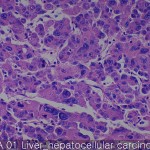
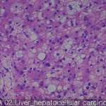
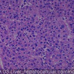
| Description | Primary cancer: 40 cores Metastatic cancer: 10 cores Non-neoplastic liver: 9 cores |
| Price | 244 EUR |
| 320 USD | |
| 210 GBP |
Product Related Literature
Liver cancer-metastasis, the disease transition stage of breast cancer that has spread to distant metastasis. Typically, the number of years you happen to resection of primary breast cancer, which is a complication of primary breast cancer. In many cases, it is possible to develop resistance to several lines of prior treatment, metastasize to distant sites them, and get a special property, metastatic cells of breast cancer, that it is very dangerous they, It is different from the properties of primary breast cancer as a recipient of the previous conditions often. That have a poor prognosis in many cases, distant metastases, accounting for about 90% of deaths from breast cancer. Bone metastatic lymph node, and lung, liver, brain, mainly breast cancer is a bone in the most common site. On several nodes and surrounding sentinel lymph node, it is considered metastatic breast cancer and local events treatable, and to be later or when it occurs in the presentation of the original lymph node metastasis.
Physical (basement membrane), chemical (ROS or reactive oxygen species, hypoxia and low pH) and biological barriers typical environment in the event of metastatic extracellular matrix and regulations (immune surveillance inhibit cytokine (ECM ) peptide) component is included. Anatomical study of organ-specific effects on the transition also includes a blood flow pattern of the primary tumor, and targeting ability of cancer cells to specific tissues. Perhaps, the targeting of cancer cells to specific organs is regulated by adhesion molecules and chemoattractant factors derived from cell surface receptors and organ specific expression on tumor cells.
Most of these processes, I need a delicate balance between the function of (TIMPs) tissue inhibitor of metalloproteinase natural matrix metalloproteinase disintegrin metalloprotease or (MMP) and (Adams). Regulatory proteolysis is an important mechanism for maintaining homeostasis. In order to provide them with the tools required to release and degradation of the extracellular matrix of the transmembrane receptors or growth factors, protease expression system in cancer cells is increased. The MMP-2, is defined in the bone, an increase in MMP-19 levels and MMP-1 was observed in the brain. In order to provide a viable cell adhesion was increased in turn, cell motility, cell migration, invasion, and growth of cancer cells, which upregulates signaling pathway.
Fibronectin is a glycoprotein of the extracellular capable of binding to ECM components other heparan sulfate proteoglycans and integrin, collagen, and fibrin, and (the HSPG). Integrin several different bind fibronectin. Signaling through the interaction of fibronectin integrin is important for cell growth migration of tumor cells, invasion and metastasis via the integrin. Adhesion of tumor cells via integrin and ECM protein tyrosine phosphorylytion nuclear translocation mitogen-activated protein activated and the focal adhesion kinase (MAP) kinase can induce regulatory signaling and gene expression Reason increased.
Heparanase, the line is disconnected to heparin sulfate HSPG you have a large network with several proteins in the ECM and cell surface. Is composed of core proteins, proteins containing fibronectin ECM various laminin, interstitial collagen, HSPG basic structure (HS) chains are covalently bound O-bound binding linear heparin sulfate some heparin-binding The lipoproteins.HSPGs, well-known components of the vessel and acts as an assembly growth factors, chemokines. Bind vascular endothelial growth factor in (VEGFs), HS stable FGF protects them from inactivation. It helps the GFS removal functions as a low-affinity co-receptor that promotes dimerization of FGF factor in circulating low concentration of growth factors, HS chains induces activation of the signal tyrosine kinase receptor further. It is expressed by cancer cells involved in angiogenesis and neovascularization deterioration of skeletal polysaccharide endothelial BM, therefore, heparanase release of angiogenic growth factors from the ECM.
ECM protein tenascin C (TNC) is regulated by metastatic breast cancer. TNC is an extracellular matrix glycoprotein that adheres adjusted. It is expressed in the tumor stroma, very has stimulated the growth of tumor cells. TNC doubt stimulated invasion through up-regulation of MMP-1 expression through activation of the MAPK pathway. Therefore (interstitial collagenase) cuts the X collagen types I, II, III, and VII, can tenascin C for the representation to change the migration of collagen influence tumor cells and ECM in cartilage tissue significantly MMP-1 You. Yield cell surface binds to co-receptor of the TGF-β for RGD and other ligands and integrin – a disulfide cross-linked homodimeric glycoprotein. Brain metastatic breast cancer cells to express a large amount of Glynn. Endoglin invasion projection to many developed endoglin overexpressing cells are localized to these structures. Leads to expression in tumor cells, I am contributing to the MMP-19 and MMP-1 to metastatic up-regulated. For example, the MMP-19 digestion basement membrane components as ECM other proteins collagen type IV, laminin 5, nidogen (entactin), fibronectin and tenascin, aggrecan and such. Thus, the yield of overexpression alters the balance of proteolytic cell for matrix degradation greatly increase the invasive properties of breast cancer.


