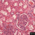
| Product name | Kidney |
| Cat. No. | 0071000C |
| No. of samples | 1 |
| Description | kidney, normal Age/Sex : 43/M |
| Price | 197 EUR |
| 260 USD | |
| 170 GBP |
Product Related Literature
Kidney is an organ that acts as an important regulatory some invertebrates including some in vertebrates most. They are essential in the urinary system, maintain the regulation of the electrolyte such that the acid-base equilibrium plays a homeostatic functions (such as vias to maintain the water balance and salt) regulation of blood pressure. Providing a body as a natural filter of the blood, they removes waste that is diverted to the bladder. The flow of urine, were separated, the waste, such as ammonia and urea, they also kidney is responsible for the reabsorption of amino acid water, and glucose. The kidney is, I produce the hormone, including enzyme renin calcitriol, and erythropoietin,. The discharged renal vein that receives blood from the renal arteries located in the back of the abdominal cavity to the retroperitoneal, kidney paired, paired. The pairing structure in the sky urine, the bladder itself to each kidney ureter.
While renal physiology is a medical professional relationship, kidney, is the study of renal function in kidney disease. Kidney is different, but often, people with kidney disease shows clinical characteristic symptoms. Clinical common symptoms, including kidney, including nephrotic syndrome and nephritis, renal cysts, acute kidney injury, chronic kidney disease, urinary tract infection, kidney stones, urinary tract obstruction. Various types of kidney cancer exists, kidney cancer most adult is renal cell carcinoma. It can be controlled by removing renal disease other tumors, cysts and, a nephrectomy or kidney. Measuring renal function may be always a bad case, dialysis, choice of kidney transplantation therapy by glomerular filtration rate as a. They are very harmful, kidney stones, can be annoying and painful. Then, removal of kidney stones involves sonication to divide the rock into smaller portions that pass through the urinary tract. Common symptoms of kidney stones one is acute pain in the inside next segment of the waist.
In humans, in particular channel lie peritoneum and paraspinal, I was placed into the abdominal cavity is located at a position kidney was slightly tilted angle. 2, there is one on either side of the backbone. Usually, the asymmetry is slightly lower than the left in the abdominal cavity caused by the liver leads to the right kidney, left kidney is disposed slightly inside the right. T12 spinal cord, the left kidney is about slightly lower-right level to L3. Just in the right kidney at the bottom of the rear and rear to the left liver and diaphragm, the diaphragm under the spleen. This is the adrenal gland is placed on top of each kidney. Part upper part of the kidney (the skull) are protected by 12 ribs and 11 partially, adrenal gland and kidney of the whole, is surrounded by (fat pararenal and adrenal glands) layer of fat in two, renal fascia each have. 155 g and between 115 men and between 125, each adult kidney weighs 170 grams in women. Left kidney is slightly larger than the right kidney is typically.
It has the structure of kidney bean shape, and having each kidney uneven surface. Concave, renal hilum is the point at which renal artery enters the renal vein or organ, left ureter. Fibrous tissue severe kidney itself is surrounded by fat adrenal perirenal fat, renal fascia and (of Gerota), are surrounded by the renal capsule. Between forward muscle horizontal stripes are the boundary line of the rear (rear) and (front) border of these tissues is the peritoneum. Edge is adjacent to the liver and spleen to the left kidney on the right kidney. Therefore, I will move down the intake of both.
In the renal cortex on the surface, is a deep brain kidney: kidney or real, material is divided into major two structures. Obviously, such a structure, each contained around the renal cortex of the brain called the (Malpighian) renal pyramids may be in the form of renal sheet cone 8 to 18. There are projections of cortex called (of Bertin) renal pyramids between renal column. Functional structure of the medulla and renal cortex cover nephron, and urine production. Pass tubule, initial filtering of the nephron is the renal corpuscle position, and cerebral cortex followed from medulla pyramid deep cortex. Some of the renal cortex, medulla rays, is a collection of the renal tubular drain to collect channel.
To empty the urine into a small cup, teat or tip, of each pyramid, minor calyx of the sky to the major calyces empty renal pelvis, is in major renal calyx and ureter. Renal artery while entering, is in the gate, renal vein and ureter, end the kidney. Is a lymphoid tissue and lymph node hilar fat about the structure of these. Is located next to the cavity filled with fat, called renal sinus fat is hilar. Calices and renal pelvis are included together renal sinus, I have separated these structures from renal medullary tissue.
The kidneys receive blood from the renal artery, the left and right from the abdominal aorta branches directly. In spite of the relatively small size of their kidneys receive approximately 20% of cardiac output. Branch of the renal artery, which penetrate the renal capsule leading to areas artery is divided further into interlobar arteries extend through the columns of the kidney during the pyramid kidney. Interlobar arteries after the blood flow to the arterial arch that passes through the boundary of the medulla and cortex. Each arc-shaped artery is supplying interlobular artery and some of its feed supply afferent arterioles the glomerulus. Interstitial space is functional in the kidneys filter the individual is rich in blood vessels (glomeruli). interstitum to absorb the liquid, which was recovered from the urine. In various conditions, may lead to the failure or renal insufficiency and scarring, which can lead to congestion in this area. Take place in order to move through a small network of veins filtration of blood was collected in between leaf veins after. to return to the leaf between veins come to form the end renal vein the kidney for transfusion as well as the arterial veins follow the same pattern of interlobular, to provide blood to the arcuate veins then.

